4028
Novel Technique for Assessment of Patellar Tracking During Weight-Bearing Knee Flexion Using Dynamic Upright MRI1FONAR Corporation, Melville, NY, United States, 2Lehigh University, Bethlehem, PA, United States
Synopsis
Despite decades of imaging research focused on the knee, little attention has been paid to dynamic, weight-bearing knee MRI emulating the physiologic condition of the joint. Here, we utilize dynamic imaging in alternating axial and sagittal planes to capture the patellofemoral joint of one volunteer performing one cycle of knee flexion/extension. Our findings indicate that it is possible to characterize patellar tracking in both axial and sagittal planes utilizing fast imaging sequences (<2 minutes) on mid-field upright MRI. Additionally, it is possible to quantify the patellofemoral kinematics on these images using patellar flexion angle and anteroposterior patellar translation.
Introduction
Despite decades of imaging research focused on the knee, relatively little attention has been paid to dynamic, weight-bearing knee MRI (1). Recently, clinicians and scientists with expertise in knee joint disorders have expressed a need for such dynamic weight-bearing knee MRI studies (2). A few studies have indeed established the relevance of weight-bearing MRI (3), and some recent investigations have explored the possibility of dynamic MRI for articulating joints (4).Patellofemoral disorders are common clinical problems and can result from abnormal patellar kinematics. It has been suggested that patellofemoral joint kinematics would be better studied utilizing dynamic weight-bearing knee MRI (5,6). Because patellofemoral joint disorders can lead to abnormal movement of the patella in both axial and sagittal planes (7), it is important to characterize normal movement of the patella in both of these planes during weight bearing flexion. Here, we demonstrate a novel technique for assessing and quantifying patellar kinematics in both axial and sagittal planes with weight-bearing dynamic MRI with knee flexion up to 45 degrees.
Methods
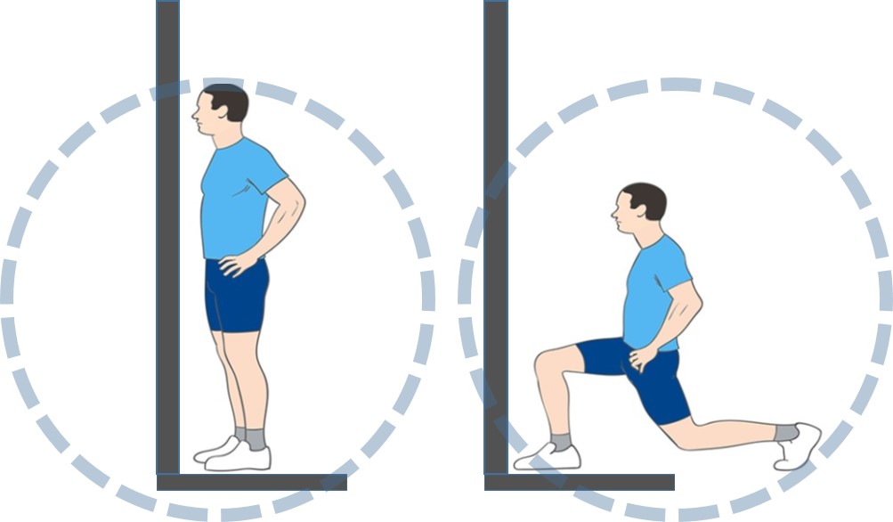
One healthy, asymptomatic volunteer underwent imaging in a 0.6T vertical gap MRI scanner where it is possible to image the patient while standing and performing weight-bearing joint movements. As shown in Figure 1, the volunteer was imaged as he performed a backward lunge, keeping the knee stable. He completed one knee flexion/extension cycle at a slow pace in approximately 40 seconds. We acquired images with a gradient echo SSFP-FID sequence, TE=3.1, TR=8.4 ms, flip angle=60 degrees, FOV=14 cm, slice thickness=5mm. The cine obtained alternating views between sagittal and axial planes. The frame time was approximately 0.8 seconds.Results
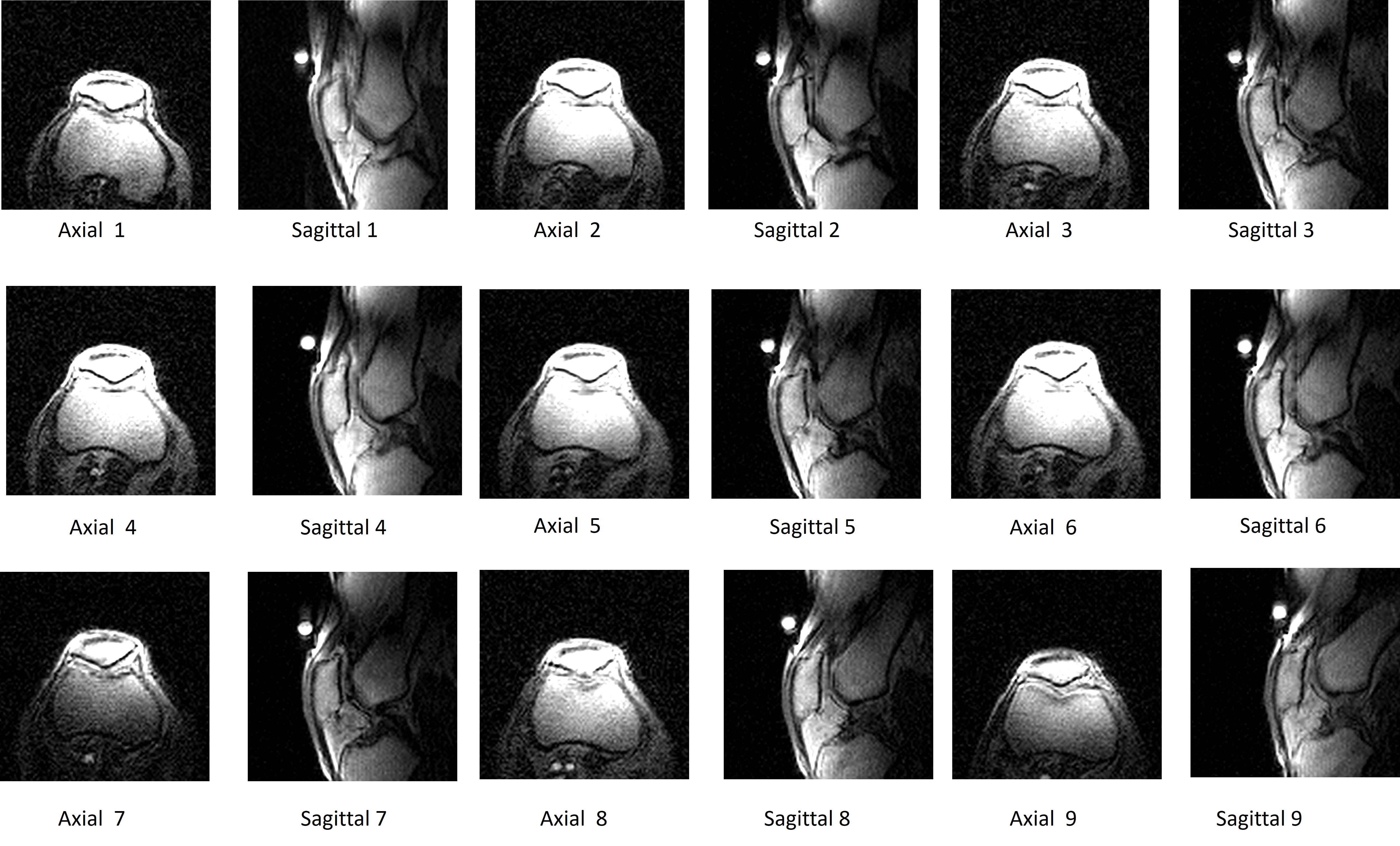
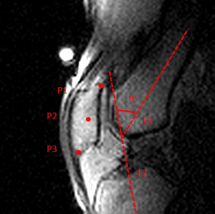
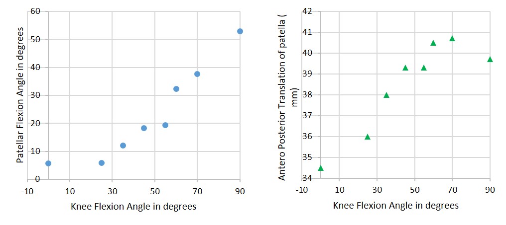
Figure 2 shows alternating images of axial and sagittal view, obtained in continuous time imaging as the knee joint underwent one cycle of weight-bearing flexion to approximately 45 degrees. Figure 3 demonstrates important anatomic landmarks for determining patellar kinematics labeled on a sagittal image. Figure 4 shows measurements of two important patellar tracking parameters: patellar flexion angle (with respect to femur) and anterior-posterior translation of patella.Discussion
Our results demonstrate preliminary proof-of-concept for conducting weight-bearing dynamic MRI of the patellofemoral joint. Qualitative findings demonstrate that it is possible to characterize patellar tracking effectively in both axial and sagittal planes simultaneously using upright MRI. We also show that it is possible to carry out measurements of patellar flexion angle and anterior-posterior translation of the patella. These patellar tracking parameters have been previously defined on radiographs, and results obtained in past studies are consistent with our findings on MRI (8).Conclusion
This case study establishes the technique needed to track patellar movement in both axial and sagittal planes utilizing fast imaging sequences (<2 minutes) on an upright MRI. Comprehensive studies using this technique are needed to examine and perfect weight-bearing MRI of the patellofemoral joint.Acknowledgements
No acknowledgement found.References
1. Jerban S, Chang EY, Du J. Magnetic resonance imaging (MRI) studies of knee joint under mechanical loading: Review. Magn Reson Imaging. 2020 Jan;65:27-36. doi: 10.1016/j.mri.2019.09.007.
2. Amano K, Li Q, Ma CB. Functional knee assessment with advanced imaging. Curr Rev Musculoskelet Med. 2016 Jun;9(2):123-9. doi: 10.1007/s12178-016-9340-0.
3. Barrance PJ, Gade V, Allen J, Cole JL. American Society of Biomechanics Clinical Biomechanics Award 2013: tibiofemoral contact location changes associated with lateral heel wedging--a weight bearing MRI study. Clin Biomech (Bristol, Avon). 2014 Nov;29(9):997-1002. doi: 10.1016/j.clinbiomech.2014.08.014.
4. Garetier, M., Borotikar, B., Makki, K. et al. Dynamic MRI for articulating joint evaluation on 1.5 T and 3.0 T scanners: setup, protocols, and real-time sequences. Insights Imaging 11, 66 (2020). https://doi.org/10.1186/s13244-020-00868-5.
5. Bruno F, Barile A, Arrigoni F, et al. Weight-bearing MRI of the knee: a review of advantages and limits. Acta Biomed. 2018;89(1-S):78-88. Published 2018 Jan 19. doi:10.23750/abm.v89i1-S.7011.
6. Yu Z, Yao J, Wang X, Xin X, Zhang K, Cai H, Fan Y, Yang B. Research Methods and Progress of Patellofemoral Joint Kinematics: A Review. J Healthc Eng. 2019 Mar 24;2019:9159267. doi: 10.1155/2019/915926.
7. PMID: 31019669; PMCID: PMC6451817.7. Nha KW, Papannagari R, Gill TJ, Van de Velde SK, Freiberg AA, Rubash HE, Li G. In vivo patellar tracking: clinical motions and patellofemoral indices. J Orthop Res. 2008 Aug;26(8):1067-74. doi: 10.1002/jor.20554.
8. Hamai S, Dunbar NJ, Moro-oka TA, Miura H, Iwamoto Y, Banks SA. Physiological sagittal plane patellar kinematics during dynamic deep knee flexion. Int Orthop. 2013 Aug;37(8):1477-82. doi: 10.1007/s00264-013-1958-6.
Figures