4020
Damping acoustic noise for MRI in Radiotherapy Settings1UMC Utrecht, Utrecht, Netherlands, 2Computational Imaging Group for MR Diagnostic & Therapy, UMC Utrecht, Utrecht, Netherlands, 3Computational Imaging Group for MR Diagnostic & Therapy, University Medical Center Utrecht, Utrecht, Netherlands
Synopsis
We present two hoods for acoustic noise damping for MRI in Radiotherapy. This is especially relevant for brain and head-and-neck patients who are scanned repetitively inside immobilization masks, which prevent wearing all the required earing protection devices. The hoods show good noise damping (average at least 10 dB attenuation) and minimal radiation dose attenuation (<1%), making them suitable also for MR-linac systems. Subjective results from a survey on 15 volunteers show that the hoods are well tolerated and do not increase significantly the feeling of claustrophobia.
Introduction
The MRI acoustic noise may induce hearing damage if protections (earplugs/earmuffs/headphones) are not properly used(1,2,3). Ideally, these devices should provide sufficient protection. This may not be the case for patients with, e.g. small ear canals, neonates, or patients in immobilization masks undergoing MRI-guided/MR-only radiotherapy(RT). Previous research proposed to use acoustic hoods as a possible solution(4,5).Here, we build upon these works and we investigate the noise attenuation for two hood designs usable in MR-simulators for MRI-guided/MR-only brain and head-and-neck radiotherapy for adult and pediatric, and potentially also in MR-linacs. First investigations on the radiation dose attenuation caused by the hoods are performed on a MR-linac. Finally, the results of a survey run on healthy volunteers to test hoods comfort during MRI are presented.
Methods
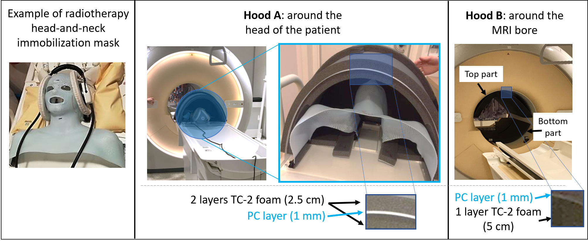
Two hoods were built to fit on the curved and flat MRI tables, and that could accommodate immobilization setups + flexible receive MR coils used for brain/head-and-neck RT(Fig.1): hood-A was made to fit around the patient head; hood-B was made to fit inside the MRI bore. For these hoods, we used TC-2 foam (Easy-Noise Control, Utrecht, NL) and Polycarbonate (supplied by WSV Kunststoffen, Utrecht, NL).Two types of acoustic noise attenuation measurements were performed:
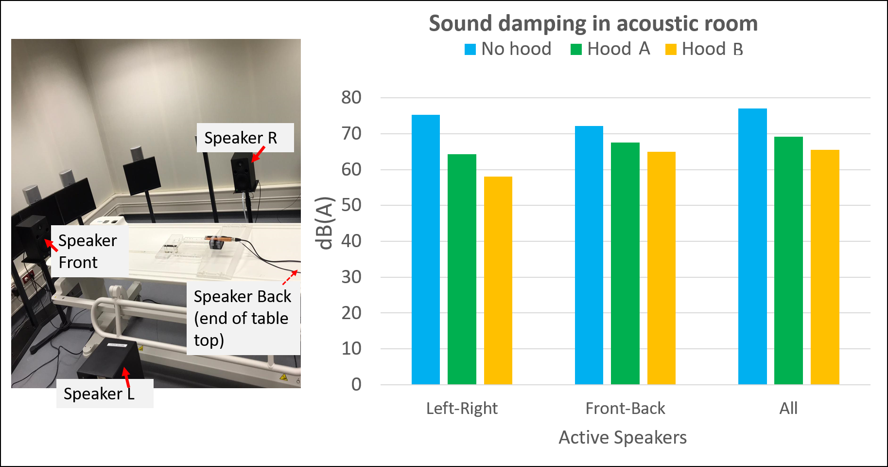
1) Inside an acoustic room (ISO-8253-2-2012) (Fig.2). Four loudspeakers(Yamaha MSP5, independently calibrated on a pink noise level of 70db(A)) were placed around the setup with a microphone at the center(Brüel & Kjær 4189 with ZC-0032 amplifier). Noise levels were recorded with the microphone and measured with a sound level meter(Brüel-Kjær 2250-S). For both hoods, three measurements were performed (feet-head speakers on; left-right speakers on; all speakers on) and compared to reference measurements without hoods.
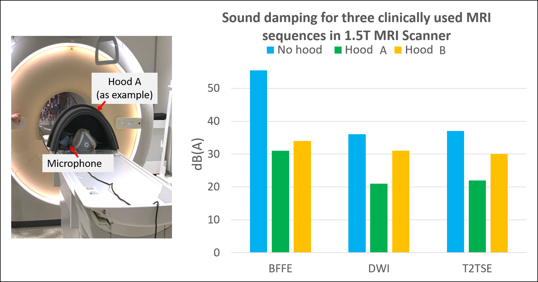
2) Inside a 1.5T MRI (Ingenia, Philips, NL) for three clinically used MRI sequences for brain RT(Fig.3). Measurements were done with a microphone (Behringer-ECM8000), recorded with Audacity and processed in Matlab. For these measurements, the measured noise is relative to the background noise intensity of the MRI room used as a reference (relative measurements).
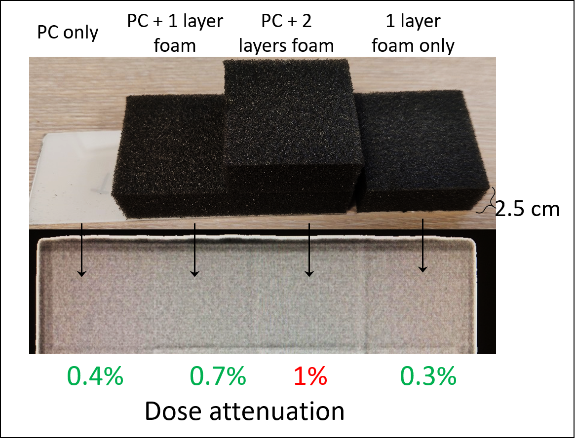
To test the beam attenuation of the hood materials, radiation measurements were performed with the megavoltage imager on the Unity MR-linac (Elekta). Relative measurements were made with and without hood materials. A correction was applied for the non-uniform dose profile of the beam.
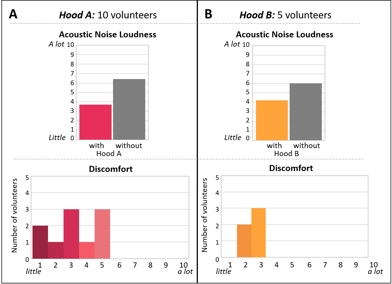
Finally, a survey was conducted on healthy volunteers (n=10 for hood-A, n=5 for hood-B) to test comfort of the developed hoods and the subjective perception of acoustic noise attenuation given by the hoods (all volunteers underwent the same MRI scan both with and without hoods).
Results
Acoustic noise attenuation measurements in the acoustic room showed noise attenuations of 8dB for hood-A and 11.5dB for hood-B (all speakers active)(Fig.2).In MRI, hood-A led to a decrease in acoustic noise between 15-25dB(Fig.3) compared to the same measurements without hood, whereas hood-B showed a decrease between 5-16dB.
Radiation dose attenuation measurements in the MR-linac showed minimal dose attenuation (0.4% for a PC layer 1 mm thick, and 0.3% for a TC-2 foam layer 2.5cm thick)(Fig.4). Achieving a dose attenuation in the range 0–1% will probably eliminate the need to apply corrections to a radiotherapy treatment plan due to the presence of the hood. These results indicate that this requirement can be satisfied.
From the survey results(Fig.5) we can observe that all volunteers reported a considerable reduction in the acoustic noise felt during MRI exams when the hoods were present, and that the presence of the hoods did not create additional discomfort.
Discussion
The presented results demonstrate experimentally the feasibility of adopting acoustic hoods to attenuate the MRI acoustic noise, especially relevant for MRI in radiotherapy settings, where patients undergo several MRI sessions in a short period of time, which may lead otherwise to hearing damage since these patients cannot wear all the necessary hearing protections due to the immobilization masks. The dose attenuation results indicate the possibility to use such hoods into MR-linacs.The variations between measurements in the acoustic and MRI rooms are most likely caused by small variations in setting up the hoods on the table top and by the difference source of noise (speakers vs MRI).
The reported subjective outcome from the volunteers indicates that the hoods are well tolerated. Only two volunteers experienced slightly more claustrophobia compared to MRI without hoods. Contrarily, for several volunteers the hoods reduced this feeling, especially for hood A, as this hood also prevents the volunteers from seeing when they are slid into the bore.
Ultimately, the in-bore ventilation is reduced by the presence of the hoods. This may lead to a sensation of increasing temperature. For hood-A, this may not be a problem as the whole body is outside the hood. For hood-B, little openings can be made in line with the ventilation openings of the MR bore.
Conclusion
Two acoustic noise damping hoods are developed for MRI in Radiotherapy. The presented results corroborated by the subjective volunteer responses highlight the importance of hearing protection, and how this can be easily achieved even in radiotherapy settings where all the standard hearing protections devices cannot be used due to immobilization masks.Acknowledgements
The authors thank T. Nguyen and T Coolen for their support during the experimental sessions at the MRI.References
1. Chao Jin, Huan Li, Xianjun Li, Miaomiao Wang, Congcong Liu, Jianxin Guo, Jian Yang. Temporary Hearing Threshold Shift in Healthy Volunteers with Hearing Protection Caused by Acoustic Noise Exposure during 3-T Multisequence MR Neuroimaging. Neuroradiology 2018, 286:602-609. doi.org/10.1148/radiol.2017161622
2. Adam Sheppard, Yu-Chen Chen, Richard Salvi. MRI Noise and Hearing Loss. The Hearing Journal 2018, 71: 30,33 doi: 10.1097/01.HJ.0000532395.75558.2d
3. Maryam Bahaloo,Mohammad Hossein Davari, Mohammad Sobhan, Seyyed Jalil Mirmohammadi,Mohammad Taghi Jalalian, Mohammad Javad Zare Sakhvidi, Farimah Shamsi, Sam Mirfendereski, Abolfazl Mollasadeghi, Amir Houshang Mehrparvar. Hearing Thresholds Changes after MRI 1.5T of Head and Neck. Radiology Research and Practice 2019: 8756579. doi: 10.1155/2019/8756579
4. Anders Nordell, Marcus Lundh, Sandra Horsch, Boubou Hallberg, Ulrika Aden, Bo Nordell, Mats Blennow. The acoustic hood: A patient-independent device improve acoustic noise protection during neonatal magnetic resonance imaging. Acta Paediatr. 2009, 98:1278-1283. doi: 10.1111/j.1651-2227.2009.01339.x.
5. Erik Huijing, Koenraad Rhebergen, Patrick Stroosnijder, Dennis. Klomp, Jannie Wijnen, Marielle Philippens, Kim Annink, Stefano Mandija..Damping of acoustic noise for fetal/neonatal MRI and MR-guided radiotherapy. ISMRM 2020: 4211.
Figures