3933
Clinical utility of turbo gradient and spin echo BLADE-DWI (TGSE-BLADE-DWI) for orbital tumors compared with readout-segmented echo-planar DWI1Department of Radiology, Union Hospital, Tongji Medical College, Huazhong University of Science and Technology, Wuhan, China, 2Hubei Province Key Laboratory of Molecular Imaging, Wuhan 430022, China, Wuhan, China, 3MR Collaboration, Siemens Healthcare Ltd., Guangzhou, China., Guangzhou, China, 4Siemens Shenzhen Magnetic Resonance Ltd., Shenzhen, China., Shenzhen, China
Synopsis
2D Turbo gradient and spin echo BLADE diffusion-weighted imaging (TGSE-BLADE-DWI) has been shown to improve image quality by reducing geometric distortions and susceptibility artifacts for middle ear cholesteatomas and optic neuritis; however, its use in depicting orbital tumors is still unknown. The results of this study demonstrated that TGSE-BLADE-DWI could provide significantly better image quality and comparable diagnostic performance for characterizing orbital tumors.
Purpose
We aimed to qualitatively and quantitatively compare the image quality and diagnostic performance of 2D turbo gradient and spin echo BLADE diffusion-weighted imaging (TGSE-BLADE-DWI) vs. readout-segmented echo-planar DWI (RESOLVE-DWI) in the evaluation of orbital tumors.Materials and methods
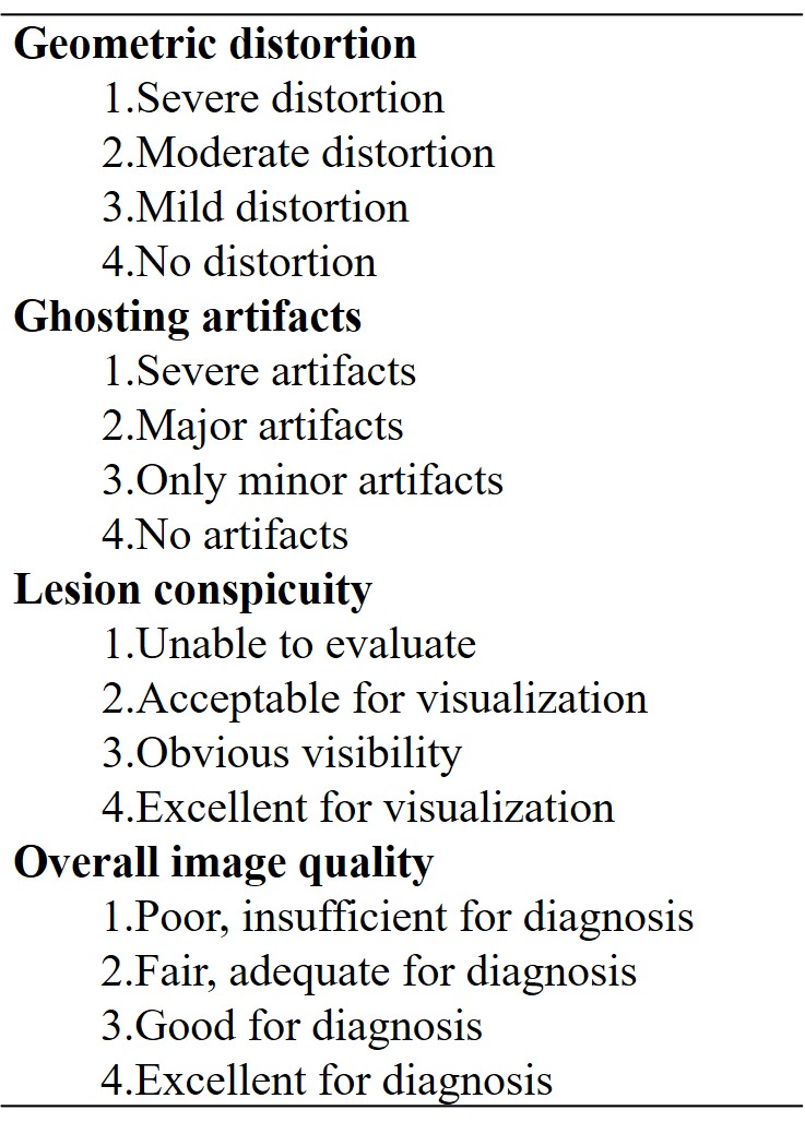
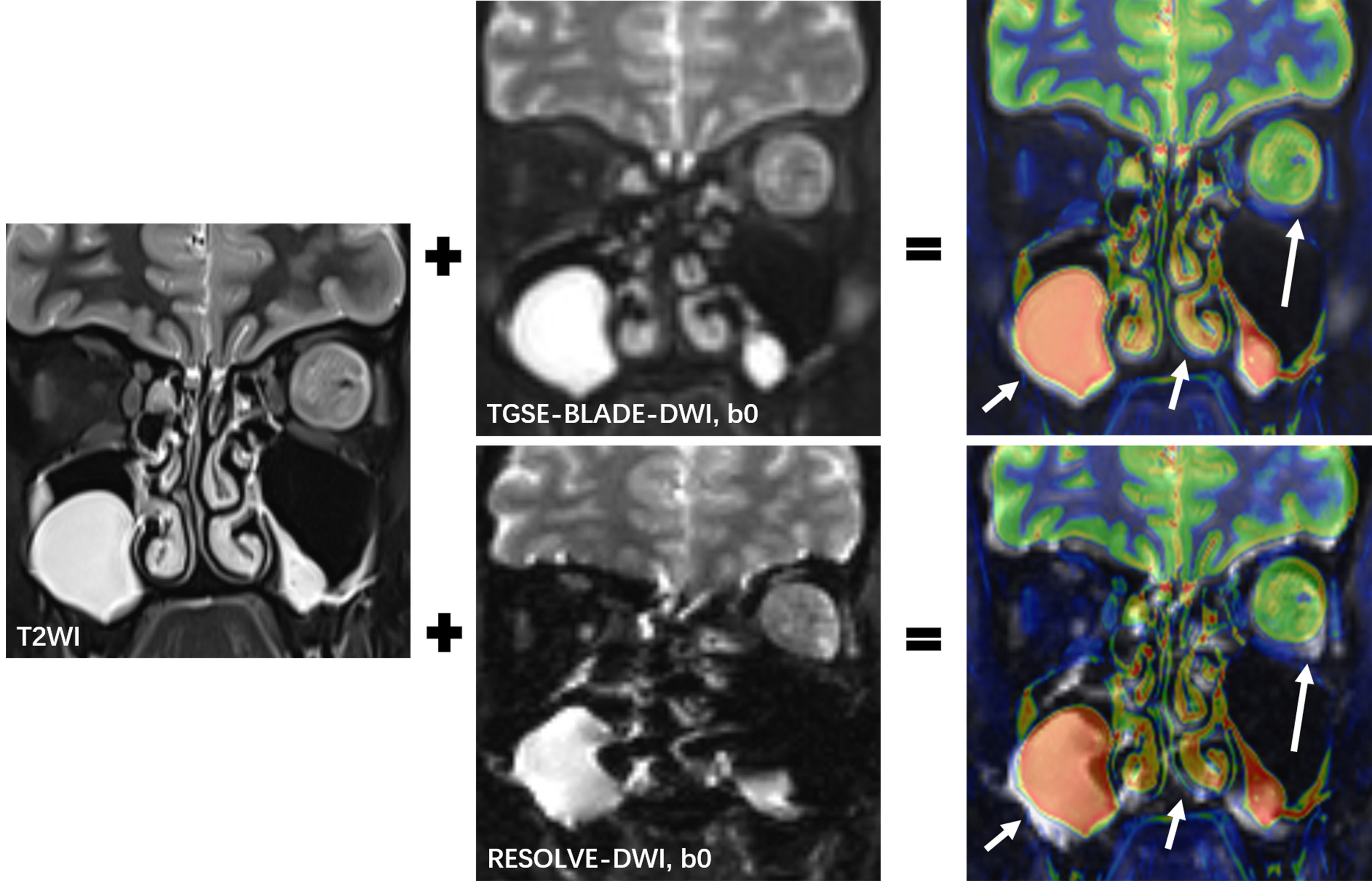
This study was approved by the local medical ethics committee, and written informed consent was obtained from each enrolled patient. Both prototypic TGSE-BLADE-DWI and RESOLVE-DWI were acquired with comparable spatial resolutions from fourteen patients with orbital tumors on a 3T MR scanner (MAGNETOM Skyra, Siemens Healthcare, Erlangen, Germany). The detailed TGSE-BLADE-DWI parameters were as follows: TR/TE: 4400/59 ms, FOV: 230 x 230 mm2, bandwidth: 650 Hz/pixel, voxel size: 1.2 x 1.2 x 2.5 mm3, number of slices: 16, matrix size: 192 x 192, BLADE coverage: 214.3%, diffusion encoding mode: 4-scan-trace, b values: 0 and 1000 s/mm2, averages: 1 for b=0 s/mm2 and 3 for b=1000 s/mm2, turbo factor: 11, EPI factor: 5, and acquisition time: 5 min 51s. The detailed parameters for RESOLVE-DWI were as follows: TR/TE: 6670/68 ms, FOV: 200 x 200 mm2, bandwidth: 650 Hz/pixel, voxel size: 1.1 x 1.1 x 2.5 mm3, number of slices: 16, matrix size: 178 x 178, diffusion encoding mode: 4-scan-trace, b values: 0 and 1000 s/mm2, averages: 1 for b=0 s/mm2 and 3 for b=1000 s/mm2, EPI factor: 89, and acquisition time: 5 min 35s.Qualitative evaluation: Two experienced radiologists scored the overall image qualities, distortions, and lesion conspicuities using a 4-point scale, as listed in Table 1. Image fusions between the T2-weighted imaging (T2WI) and two DWI sequences (b=0 s/mm2) were used as references for grading the extent of geometric distortions (Fig 1).
Quantitative evaluation: Quantitative measurements, including contrast-to-noise ratios (CNRs), and apparent diffusion coefficients (ADCs) of orbital tumors, and geometric distortion rates (GDRs) of the two DWI sequences were calculated and compared.
CNR=(SIlesion-SIWM)/√[(SDlesion)2+(SDWM)2]
SIlesion and SIWM represent the mean signal intensities of lesions and white matter. SDlesion and SDWM represent the standard deviation of lesions and white matter. All measurements were performed, avoiding signals from adjacent air, bone, and susceptibility artifacts. The GDR was calculated as follows:
GDR= (LT2 -LDWI)/ LT2 ×100%
LT2 and LDWI represent the maximum length of the vitreous body.
Results
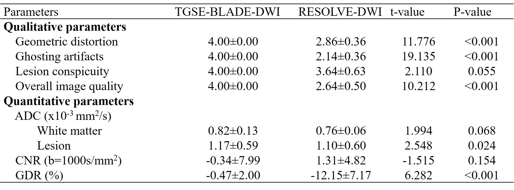
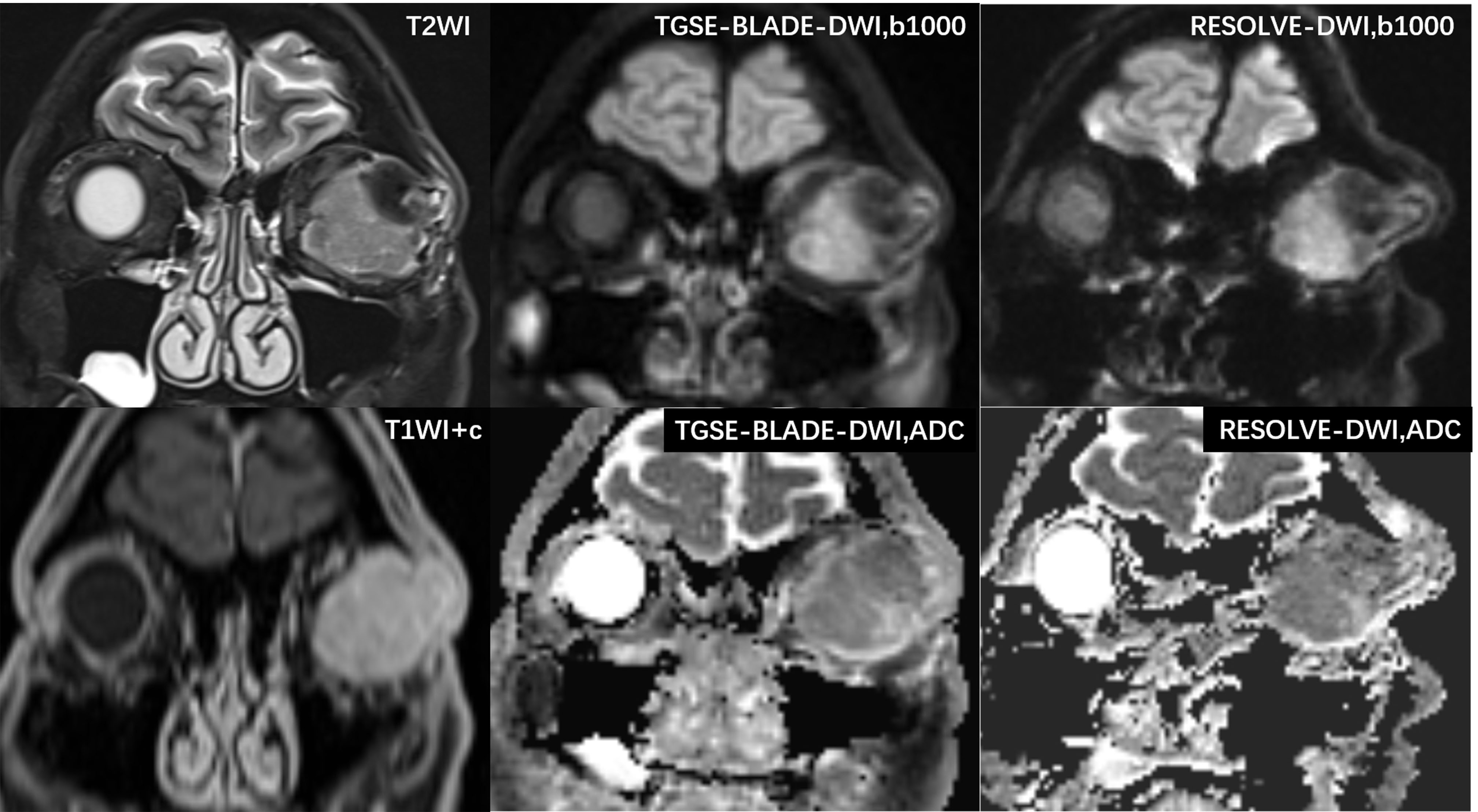
Qualitative and Quantitative evaluation results are displayed in Table 2. There was no significant difference in the lesion conspicuity scores between the two DWI sequences (p=0.055). In contrast, TGSE-BLADE-DWI scored higher than RESOLVE-DWI for geometric distortions, ghosting artifacts, and overall image quality (all p <0.01).For quantitative GDR evaluations, TGSE-BLADE-DWI showed reduced geometric distortions compared with RESOLVE-DWI (p<0.01) (Fig 1, arrows). There was no significant difference in orbital tumor CNRs and white matter ADCs between the two DWI sequences (p=0.154, p=0.068, respectively). ADC values of TGSE-BLADE-DWI was higher than those of RESOLVE-DWI (p=0.024). Figure 2 showed that left orbital tumors could be visualized clearly using both DWI sequences, and the TGSE-BLADE-DWI showed improved distortions and less ghosting artifacts compared to RESOLVE-DWI. The ADC values of this lesion on TGSE-BLADE-DWI and RESOLVE-DWI were 1.24 x 10⁻³ mm2/s and 1.11 x 10⁻³ mm2/s.
Conclusions
Compared with RESOLVE-DWI, TGSE-BLADE-DWI with reduced geometric distortion and ghosting artifacts significantly improved image quality for the evaluation of orbital tumors.Acknowledgements
Not applicable.References
1. Xu X, Wang Y, Hu H, Su G, Liu H (2017) Readout-segmented echo-planar diffusion-weighted imaging in the assessment of orbital tumors: comparison with conventional single-shot echo-planar imaging in image quality and diagnostic performance. Acta Radiol 58:1457–1467.
2. Kammer NN, Coppenrath E, Treitl KM, et al (2016) Comparison of contrast-enhanced modified T1-weighted 3D TSE black-blood and 3D MP-RAGE sequences for the detection of cerebral metastases and brain tumours. Eur Radiol 26:1818–1825.
3. Zhou K, Liu W, Cheng S (2018) Non-CMPG PROPELLER diffusion imaging: comparison of phase insensitive preparation with split acquisition. In: Proceedings of the Annual Meeting of the International Society for Magnetic Resonance in Medicine;5320.
4. Sheng Y, Hong R, Sha Y, et al (2020) Performance of TGSE BLADE DWI compared with RESOLVE DWI in the diagnosis of cholesteatoma. BMC Med Imaging 20:40.
5. Yuan T, Sha Y, Zhang Z, et al (2020) TGSE diffusion-weighted pulse sequence in the evaluation of optic neuritis: A comprehensive comparison of image quality with RESOLVE. Proc. Intl. Soc. Mag. Reson. Med. 28.
6. Xu X-Q, Liu J, Hu H, et al (2016) Improve the image quality of orbital 3 T diffusion-weighted magnetic resonance imaging with readout-segmented echo-planar imaging. Clinical Imaging 40:793–796.
Figures