3918
Development of MR Elastography Methods for Assessing Adhesion Between Pituitary Masses and the Optic Chiasm1Radiology, Mayo Clinic, Rochester, MN, United States, 2Neurologic Surgery, Mayo Clinic, Rochester, MN, United States
Synopsis
Preoperatively knowing if a pituitary adenoma is adhering to the optic chiasm would help to predict the risk of surgically induced vision loss. MR-elastography based slip interface imaging has been successfully developed to predict tumor adhesion using normalized octahedral shear strain (NOSS). This pilot study demonstrates the feasibility of acquiring NOSS at ~1mm high resolution using a newly developed distortion-free EPI-MRE technique on a high-performance compact 3T system. With further development, this technique will have great potential to identify adhesion between pituitary adenomas with the adjacent optic chiasm.
Introduction
MR elastography (MRE) has become a useful tool to reliably characterize tumor consistency and adherence, offering key information for surgical planning.[1, 2] For pituitary adenomas, preoperatively knowing if the tumor is adhering to the optic chiasm would help to predict the risk of surgically induced vision loss. Slip interface imaging using spin-echo (SE) EPI-MRE has been successfully developed to predict tumor adhesion using normalized octahedral shear strain (NOSS) [3]. However, the sella containing the pituitary adenoma is adjacent to the sphenoid sinus which is associated with susceptibility artifacts, and the optic chiasm is only 2 to 3 mm thick and may be stretched thin over the tumor. These challenges make it substantially difficult to image the interface with the standard SE-EPI-MRE technique that typically acquires data at 2-3mm resolution and is prone to image distortion. Recently, we have developed a distortion-free SE-EPI-MRE technique (that we refer to as DIADEM-MRE) [4], based on distortion-free EPI technologies [5-7] and demonstrated its application for high-resolution brain MRE on a high-performance compact 3T MRI. In this study, we further developed the technique to achieve ~1mm in-plane resolution for better coronal plane imaging of the adenoma-nerve interface and demonstrated its feasibility on 3 pituitary adenoma patients.Methods
The distortion-free DIADEM-MRE technique is based on a multi-band spin-echo EPI-MRE sequence [8] with an additional spin-warp phase-encoding gradient immediately before the EPI acquisition [9, 10]. With IRB approval and written informed consent, 3 pituitary adenoma patients were scanned on the compact 3T scanner [11-13] capable of achieving simultaneous gradient amplitude of 80mT/m and slew rate of 700T/m/s with negligible peripheral nerve stimulation [14], using a Nova Medical 32-channel receiver coil. DIADEM-MRE was performed in the coronal plane with 1.1-mm resolution in the readout direction (across the chiasm in the superior-inferior direction) and 2-mm resolution in the phase and slice directions. The imaging parameters were TR/TE=1700/69.1ms; FOV=22 cm; 192×110 acquisition matrix, reconstructed to 440x440; 34 contiguous 2-mm-thick coronal slices; 2x phase acceleration; 2x multiband (MB) acceleration, 464 μs echo spacing, 60-Hz mechanical vibrations; 6 motion encoding directions, 3 phase offsets; 11 shots for each DIADEM dataset and a total acquisition time of 5:40 minutes. For the NOSS calculation, first the phase difference images were unwrapped with a previously described dual-motion-sensitivity encoding method [15, 16], and were then converted to 3D displacement fields. Next the shear wave fields were calculated by removing the rigid body motion from the 3D displacement fields using rigid body fitting [15]. Finally, NOSS was calculated from the shear wave fields using the method described in the previous study [3].Results
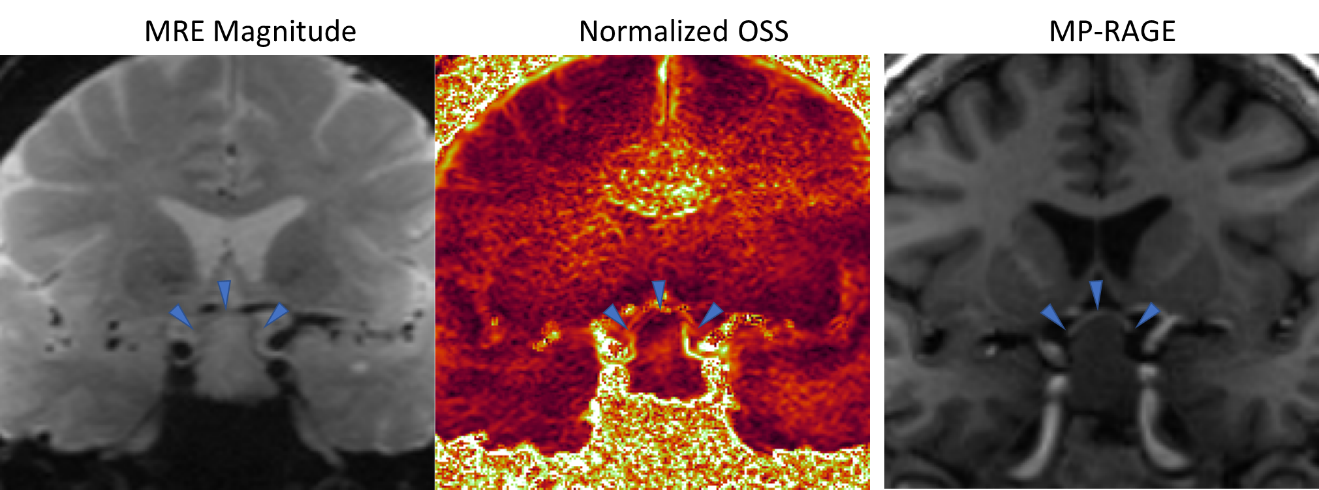
Figure 1 shows the results from one patient with a non-functioning pituitary adenoma who underwent an endoscopic endonasal approach for tumor resection. During surgery, no tumor adhesion was noted to the optic apparatus. The distortion-free EPI-MRE magnitude, NOSS map, and MP-RAGE image were co-registered to each other. The thin optic chiasm is visible on the high-resolution MRE magnitude images (indicated by blue triangles). Two high-intensity normalized OSS lines can be seen on both sides of the nerve, which would be consistent with a non-adhesive interface.Discussion and Conclusion
This preliminary result demonstrates the feasibility to acquire NOSS at ~1mm resolution using DIADEM-MRE on a high-performance compact 3T system. The distortion-free MRE images make it possible to identify the optic chiasm by directly comparing with the anatomical images without any EPI distortion correction that may compromise the image resolution. Although the 3 patients imaged to date had no reported adhesion, this pilot study demonstrates great potential to identify adhesion between pituitary adenomas with the adjacent optic chiasm.Acknowledgements
This work was supported by grants from the NIH (R01 EB001981, R01 NS113760, and U01EB02445) and Mayo Clinic imaging awards CIM-92541587.
References
1. Murphy, M.C., et al., Preoperative assessment of meningioma stiffness using magnetic resonance elastography. Journal of Neurosurgery, 2013. 118(3): p. 643-648.
2. Yin, Z., et al., Slip interface imaging predicts tumor-brain adhesion in vestibular schwannomas. Radiology, 2015. 277(2): p. 507-517.
3. Yin, Z., et al., Slip interface imaging based on MR-elastography preoperatively predicts meningioma-brain adhesion. J Magn Reson Imaging, 2017. 46(4): p. 1007-1016.
4. Sui, Y., et al. High-Resolution Distortion-Free Whole-Brain MR Elastography using Multiband DIADEM (DIADEM-MRE). in ISMRM. 2020.
5. In, M.H., et al., Distortion-free imaging: A double encoding method (DIADEM) combined with multiband imaging for rapid distortion-free high-resolution diffusion imaging on a compact 3T with high-performance gradients. J Magn Reson Imaging, 2019.
6. In, M.H., O. Posnansky, and O. Speck, High-resolution distortion-free diffusion imaging using hybrid spin-warp and echo-planar PSF-encoding approach. Neuroimage, 2017. 148: p. 20-30.
7. Dong, Z., et al., Tilted-CAIPI for highly accelerated distortion-free EPI with point spread function (PSF) encoding. Magn Reson Med, 2019. 81(1): p. 377-392.
8. Sui, Y., et al., Fast Brain MR Elastography Using a Simultaneous Multislice EPI Acquisition on a Compact 3T Scanner. ISMRM Proceeding, 2019: p. 3971.
9. Robson, M.D., J.C. Gore, and R.T. Constable, Measurement of the point spread function in MRI using constant time imaging. Magnetic resonance in medicine, 1997. 38(5): p. 733-740.
10. Zeng, H. and R.T. Constable, Image distortion correction in EPI: comparison of field mapping with point spread function mapping. Magn Reson Med, 2002. 48(1): p. 137-46.
11. Weavers, P.T., et al., Technical Note: Compact three-tesla magnetic resonance imager with high-performance gradients passes ACR image quality and acoustic noise tests. Med Phys, 2016. 43(3): p. 1259-1264.
12. Foo, T.K.F., et al., Lightweight, compact, and high-performance 3T MR system for imaging the brain and extremities. Magnetic Resonance in Medicine, 2018. 80(5): p. 2232-2245.
13. Tao, S.Z., et al., Gradient Pre-Emphasis to Counteract First-Order Concomitant Fields on Asymmetric MRI Gradient Systems. Magnetic Resonance in Medicine, 2017. 77(6): p. 2250-2262.
14. In, M.H., et al., Reducing PNS with minimal performance penalties via simple pulse sequence modifications on a high-performance compact 3T scanner. Phys Med Biol, 2020. 65(15): p. 15NT02.
15. Yin, Z., et al., In vivo characterization of 3D skull and brain motion during dynamic head vibration using magnetic resonance elastography. Magn Reson Med, 2018.
16. Sui, Y., et al. Accurate High Dynamic Range Displacement Measurement in MR Elastography Using a Simultaneous Dual-Sensitivity Acquisition. in ISMRM. 2018. Paris, France.
Figures