3915
Evaluation of Ultrafast Post-Contrast Wave-CAIPI 3D-T1 MPRAGE Compared to Standard 3D-T1 MPRAGE for Evaluating Enhancing Lesions on 3T MRI.1Department of Radiology, Massachusetts General Hospital, Boston, MA, United States, 2Department of Radiology, Athinoula A. Martinos Center for Biomedical Imaging, Charlestown, MA, United States, 3Department of Radiology, Siriraj Hospital, Bangkok, Thailand, 4Siemens Shenzhen Magnetic Resonance Ltd., Shenzhen, China, 5Siemens Healthcare GmbH, Erlangen, Germany, 6Siemens Medical Solutions Inc., Boston, MA, United States
Synopsis
We compared a highly accelerated post-contrast Wave-CAIPI 3D T1 MPRAGE sequence to the standard 3D T1 MPRAGE in 80 patients undergoing imaging evaluation of intracranial enhancing lesions on 3T MRI. Two neuroradiologists performed a head-to-head analysis to assess the images and found that post-contrast 3D Wave-T1 MPRAGE was up to 2x faster than the standard sequence with no significant difference in the detection of enhancing lesions. The application of highly accelerated Wave-T1 MPRAGE may improve use of MRI resources while reducing patient anxiety and being less prone to motion artifacts.
Introduction
Three-dimensional (3D) post-contrast imaging of the brain is essential for complete evaluation of a wide range of inflammatory, neoplastic, and neurovascular diseases of the CNS. 3D T1-weighted post-contrast sequences provide better special representation of enhancing pathology and allow precise planning for biopsies and other interventions. The inversion recovery fast gradient recalled-echo MPRAGE is the most widely used T1 post-contrast sequence and has been recommended for use in brain tumor clinical trials in recent consensus recommendations for brain tumor imaging in clinical trials1,2. However, high-resolution volumetric sequences have prolonged scanning times that may compromise image quality. The purpose of this study was to investigate the diagnostic performance of an accelerated post-contrast Wave-CAIPI T1 MPRAGE (Wave-T1 MPRAGE) sequence compared to a standard post-contrast T1 MPRAGE sequence for detection of intracranial enhancing lesions.Methods
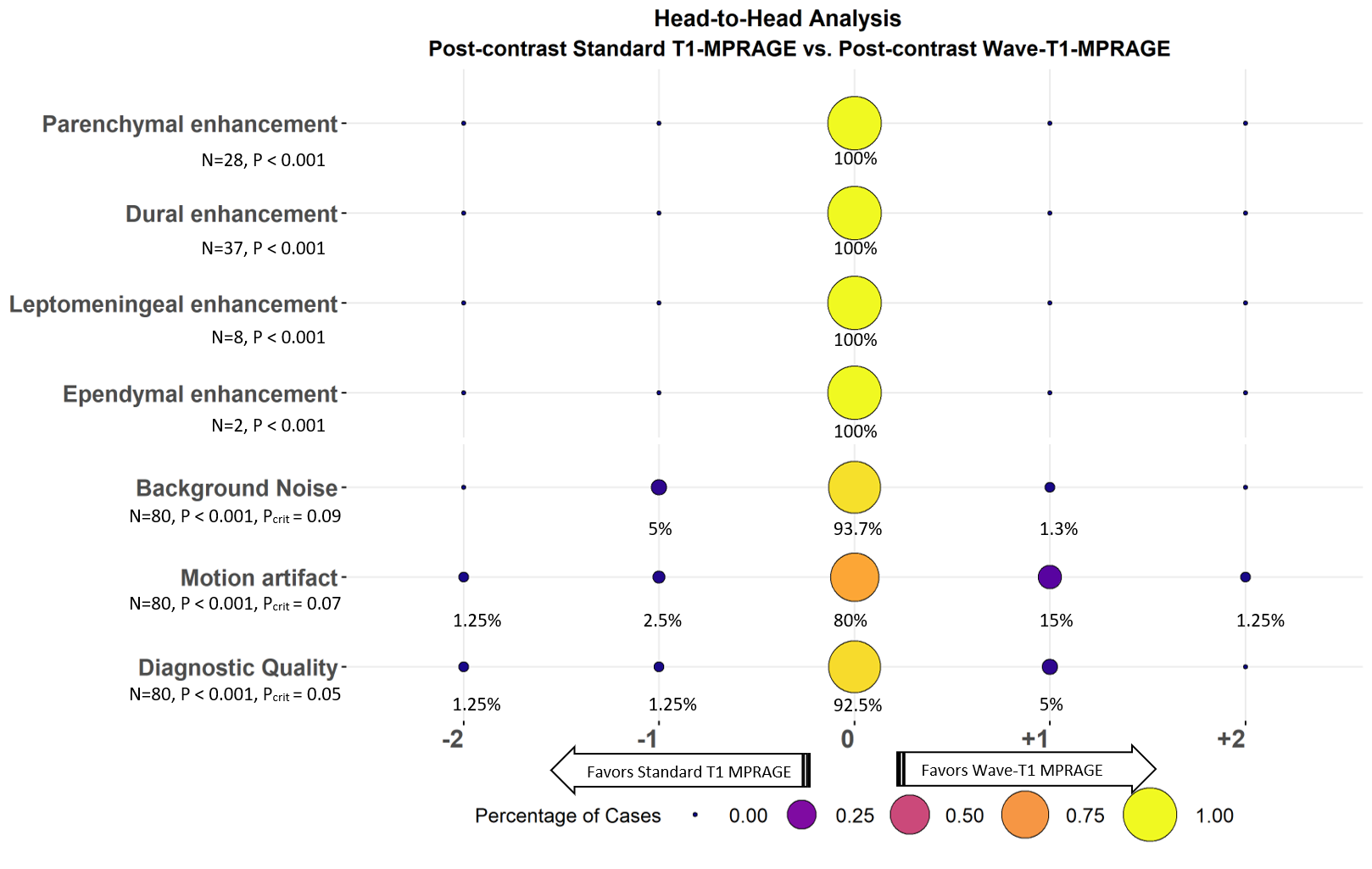
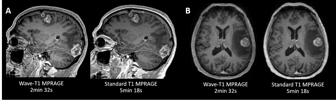
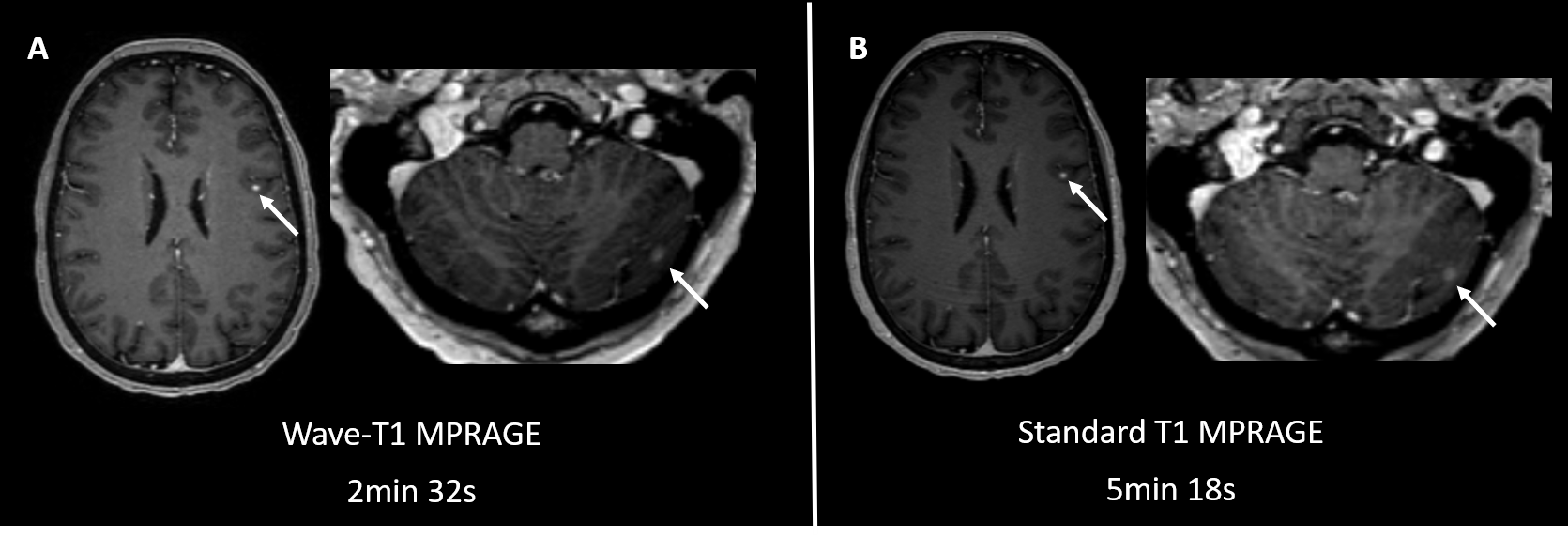
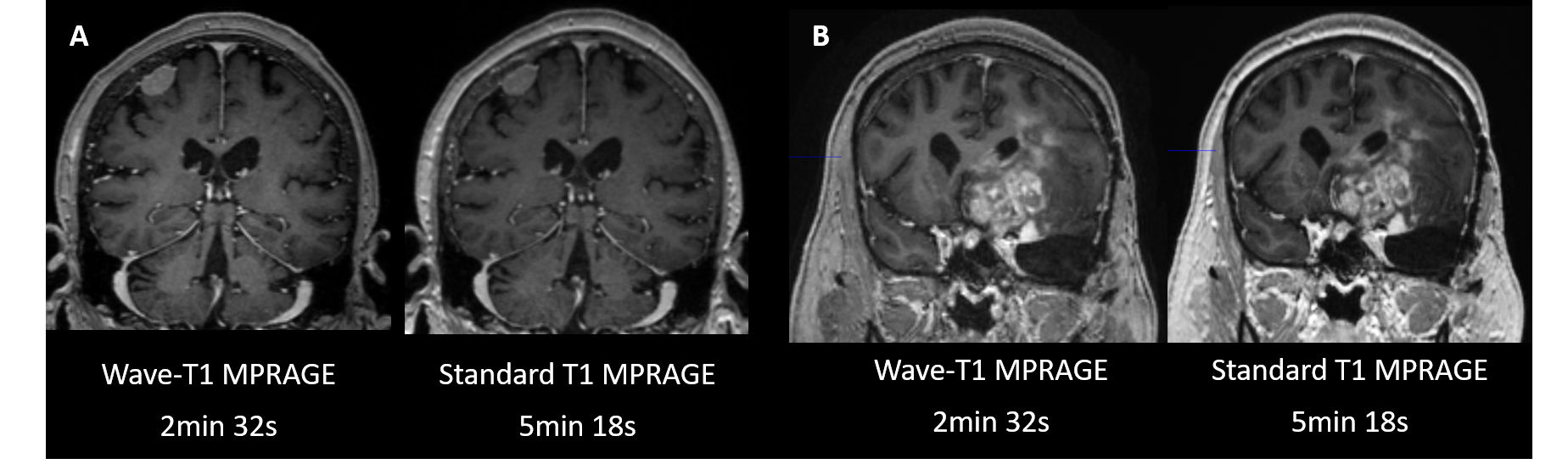
The study was approved by the institution’s IRB and was HIPAA compliant. We prospectively enrolled 80 adult patients undergoing brain MRI with contrast. The imaging protocol of all MRI scans included a standard post-contrast T1 MPRAGE (acceleration factor [R]=2, acquisition time [TA]=5min18s, 0.9 mm isotropic resolution) and a resolution-matched prototype post-contrast Wave-T1 MPRAGE sequence (R=4, TA=2min32s). 54 cases were acquired with the standard sequence before Wave, and 26 cases were acquired with Wave before standard. The studies were performed on a 3T MRI system (MAGNETOM Prisma, Siemens Healthcare, Erlangen, Germany). Two neuroradiologists (9 and 13 years of experience) blinded to sequence type performed an independent head-to-head comparison of the images. A predefined 5-point scale was used for grading parenchymal, dural, leptomeningeal, and ependymal enhancement. The raters also scored the perception of motion artifacts, noise and the overall diagnostic quality. Discrepancies among raters were adjudicated by a third reader (>20 years of experience). We performed a non-inferiority test with a 15% margin to assess the proportion of cases where the post-contrast standard sequence was preferred over Wave-T1 MPRAGE in the head-to-head analysis.Results
The evaluation of neoplasms (64%) was the most frequent indication for MRI, followed by vascular (10%) and inflammatory conditions (2%). Wave-T1 MPRAGE was equivalent to the standard sequence for detection of intracranial enhancing lesions in all locations, with a 2-fold reduction in acquisition time (2:32 vs 5:18). Wave-T1 MPRAGE images were non-inferior to the standard images in the perception of noise and motion artifacts (P<0.001). Moreover, Wave-T1 MPRAGE presented less motion artifacts in 16% of cases, showing better performance compared to the standard sequence. The diagnostic quality of the standard and Wave sequences was also equivalent (p<0.001).Discussion
Wave-CAIPI technology enables the acquisition of high-resolution 3D post-contrast T1 MPRAGE images in less than half the acquisition time3, with equivalent diagnostic performance as a resolution-matched standard post-contrast T1 MPRAGE sequence. The faster scan has the added benefit of potentially reducing artifacts due to motion, particularly in motion-prone patients. The results encourage the replacement of standard T1 MPRAGE for the Wave-accelerated T1 MPRAGE sequence in clinical brain MRI protocols with contrast. The reduction in scan time and increased scan throughput will increase patient access and improve the utilization of MR imaging resources.Conclusion
Post-contrast Wave-T1 MPRAGE provided high-resolution 3D examination of the brain with non-inferior diagnostic performance compared to the standard sequence. The clinical application of the Wave-CAIPI approach to post-contrast T1 MPRAGE may enable more efficient utilization of MR resources. Therefore, the deployment of an ultrafast Wave-T1 MPRAGE can improve efficiency in diagnosis while preserving clinically important information.Acknowledgements
This study was supported by a research grant from Siemens Healthineers.References
1. Kaufmann TJ, Smits M, Boxerman J, et al. Consensus recommendations for a standardized brain tumor imaging protocol for clinical trials in brain metastases. Neuro Oncol. 2020;22(6):757-772. doi:10.1093/neuonc/noaa030
2. Ellingson BM, Wen PY, Cloughesy TF. Modified Criteria for Radiographic Response Assessment in Glioblastoma Clinical Trials. Neurotherapeutics. 2017;14(2):307-320. doi:10.1007/s13311-016-0507-6
3. Polak D, Setsompop K, Cauley SF, et al. Wave-CAIPI for highly accelerated MP-RAGE imaging. Magn Reson Med. 2018;79(1):401-406. doi:10.1002/mrm.26649
Figures