3914
Contrast-enhanced T1-weighted DANTE-SPACE, PETRA, and MPRAGE: clinical evaluation comparison in intracranial tumor patients at 3T1Department of Radiology, Union Hospital, Tongji Medical College, Huazhong University of Science and Technology, Wuhan, China, 2Hubei Province Key Laboratory of Molecular Imaging, Wuhan, China, 3Centers for Biomedical Engineering, University of Science and Technology of China, Hefei, China, 4MR Collaboration, Siemens Healthcare Ltd., Guangzhou, China, 5Siemens Medical Solutions USA, Inc., Portland, OR, United States
Synopsis
Three dimensional (3D) T1-weighted sequences are commonly used for imaging of contrast-enhanced brain tumors, but enhancement of small vessels may mimic imaging of focal lesions, thereby hindering accurate diagnosis. This study validated that when compared with PETRA and conventional MPRAGE, DANTE-SPACE detects more small metastases with better CNR between lesions and surrounding parenchyma, and may be useful for differential tumor diagnosis because of its blood signal suppression abilities; PETRA could achieve similar detection for brain tumors and quieter acoustic noise level when compared with MPRAGE.
Purpose
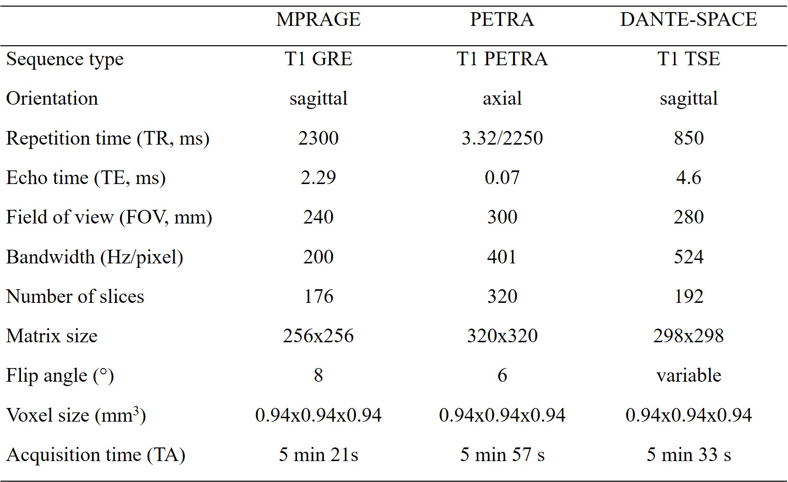
To compare the performance of three contrast-enhanced T1-weighted three-dimensional magnetic resonance sequences for detection of brain tumors at 3T: turbo spin echo imaging with variable flip angle echo train with delay alternating with nutation for tailored excitation (DANTE-SPACE), pointwise encoding time reduction with radial acquisition (PETRA), and conventional magnetization-prepared rapid gradient echo (MPRAGE).Materials and methods
The study protocol was approved by the local medical ethics committee. A total of 77 consecutive patients (43 males and 34 females; mean age, 51.6 years; age range, 20-73 years), 43 patients with lung cancer or breast cancer and 34 patients with identified primary brain tumors were enrolled. All MR examinations were performed on a 3T MR system (MAGNETOM Skyra, Siemens Healthcare, Erlangen, Germany) using a 20-channel head-neck coil. All MR sequences were 3D acquisitions covering the whole brain and were designed to acquire at the same spatial resolution with comparable acquisition times (Table 1).Signal intensities (SI) of gray matter (GM), white matter (WM), and detected lesions were measured for calculating signal-to-noise ratios (SNRs) and contrast-to-noise ratios (CNRs). SNRs for GM and WM, and CNRs for lesion to WM, lesion to GM, and WM to GM for MPRAGE, PETRA, and the prototype DANTE-SPACE sequences were calculated using the following formulas:
SNRWM=SIWM/SDWM
SNRGM=SIGM/SDGM
CNRlesion/WM= (SIlesion-SIWM )/SDWM
CNRlesion/GM= (SIlesion-SIGM )/SDGM
SD represents standard deviation of the respective tissue (GM/WM), indicative of the noise level. For CNRlesion/WM and CNRlesion/GM evaluation, lesions with homogeneous solid enhancement and clear margin and with diameter ≥5mm or larger were selected. Circular ROIs of enhancing lesions were placed in the center of the lesions without involving the margin of lesions.
Two radiologists scored the overall image quality and lesion depiction by using a 4-point scale (4, excellent, no artefacts; 3, good, minor artefacts; 2, adequate, major artifacts; 1, insufficient for diagnosis). The acoustic levels of the three sequences were recorded using a decibel meter placed laterally 1-meter from the lateral panel of the MR scanner core.
Statistical analysis: Kruskal-Wallis one-way ANOVA and Bonferroni correction were used. Intraclass correlation coefficients (ICCs) between the two radiologists for presence of metastatic lesions were assessed for inter-observer agreement.
Results
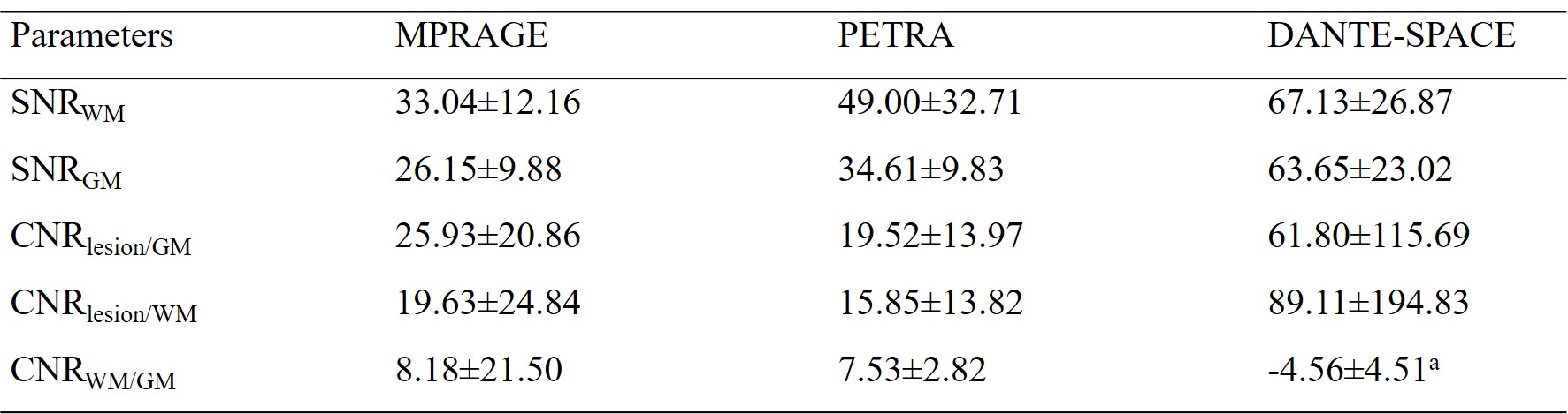
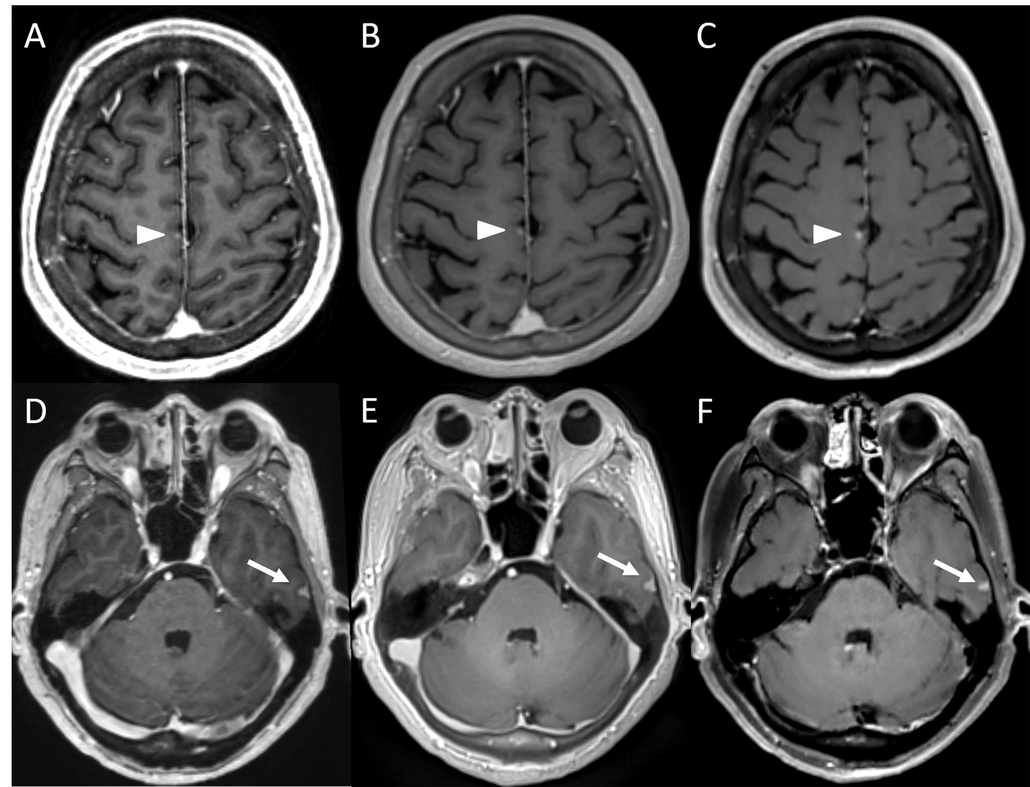
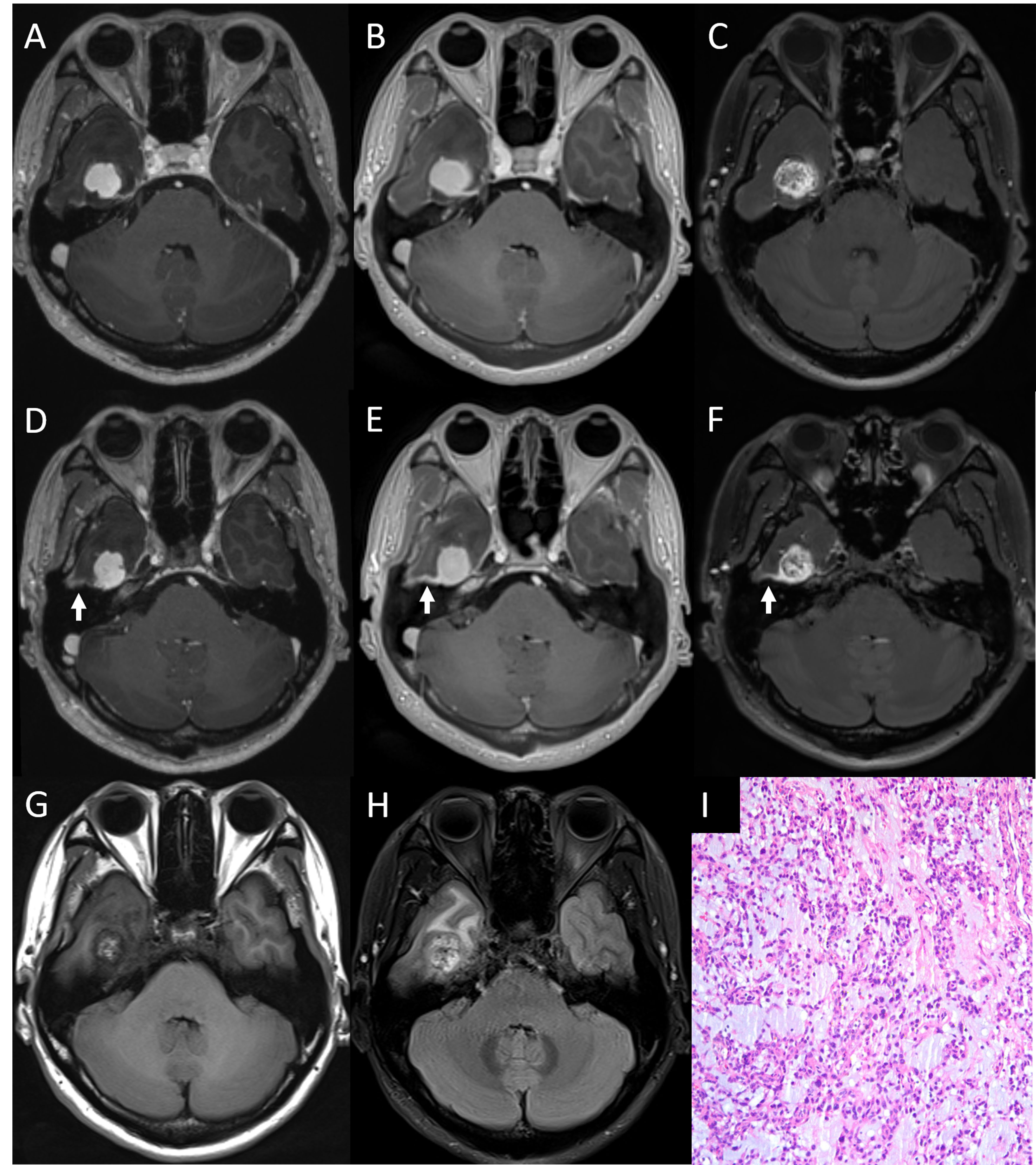
The mean (± standard deviation) values of objective parameters SNRWM, SNRGM, CNRlesion/GM, CNRlesion/WM, and CNRWM/GM across the three sequences are listed in Table 2. DANTE-SPACE SNRWM and SNRGM were significantly higher than those of PETRA (p<0.001, p<0.001, respectively), while these values in PETRA were significantly higher than those of MPRAGE (p<0.001, p<0.001, respectively). DANTE-SPACE CNRlesion/GM and CNRlesion/WM were significantly higher than those of MPRAGE and PETRA (p<0.001, p<0.001, respectively) (Fig. 1). The image scores which focused on lesion depiction for the three sequences were 3.76±0.43 (PETRA), 3.75±0.44 (MPRAGE), and 3.96±0.20 (DANTE-SPACE), with DANTE-SPACE significantly higher than that of PETRA and MPRAGE (p=0.002, p=0.004, respectively).Significantly more small brain metastases were detected with DANTE-SPACE (94 lesions) than those with MPRAGE (71 lesions) and PETRA (72 lesions). ICCs between the two radiologists for presence of metastatic lesions were 0.964 (MPRAGE), 0.975 (PETRA) and 0.973 (DANTE-SPACE). For depiction of primary tumors, no significant difference was observed between the number (n=22, n=22, n=22, p=1.000) and maximum diameter of primary brain tumors (3.45±1.87 cm, 3.49±1.85 cm, 3.49±1.85 cm, p=0.998) across sequences. However, the homogeneity of signal intensity within some tumors differed among the three sequences (Fig. 2).
The acoustic noise level of PETRA sequence (64.45±1.52 dB) was significantly lower than that of MPRAGE (78.27±2.18 dB) (p<0.01), and noise level of MPRAGE was significantly lower than that of DANTE-SPACE (80.18±1.10 dB) (p<0.01).
Conclusion
Compared to MPRAGE and PETRA, DANTE-SPACE with blood vessel suppression shows improved detection of small cerebral metastases and increased CNR between lesions and surrounding parenchyma. DANTE-SPACE may be helpful for tumor differential diagnosis.Acknowledgements
Not applicable.References
1. Mugler, J. P.; Brookeman, J. R. Three-Dimensional Magnetization-Prepared Rapid Gradient-Echo Imaging (3D MP RAGE). Magn. Reson. Med. 1990, 15 (1), 152–157.
2. Grodzki, D. M.; Jakob, P. M.; Heismann, B. Ultrashort Echo Time Imaging Using Pointwise Encoding Time Reduction with Radial Acquisition (PETRA). Magn. Reson. Med. 2012, 67 (2), 510–518.
3. Ida, M.; Wakayama, T.; Nielsen, M. L.; Abe, T.; Grodzki, D. M. Quiet T1-Weighted Imaging Using PETRA: Initial Clinical Evaluation in Intracranial Tumor Patients: Quiet T1-Weighted Imaging Using PETRA. J. Magn. Reson. Imaging 2015, 41 (2), 447–453.
4. Kim, D.; Heo, Y. J.; Jeong, H. W.; Baek, J. W.; Han, J.-Y.; Lee, J. Y.; Jin, S.-C.; Baek, H. J. Usefulness of the Delay Alternating with Nutation for Tailored Excitation Pulse with T1-Weighted Sampling Perfection with Application-Optimized Contrasts Using Different Flip Angle Evolution in the Detection of Cerebral Metastases: Comparison with MPRAGE Imaging. AJNR Am J Neuroradiol 2019, ajnr.A6158.
Figures