3900
Development and validation of radiomics model for diagnosing PCar and BPH based on Diffusion weighted imaging and clinical information
Lihua Chen1, Ailian Liu1, Yan Guo2, and Xin Li2
1The First Affiliated Hospital of DaLian Medical University, Dalian, China, 2GE Healthcare, China, Beijing, China
1The First Affiliated Hospital of DaLian Medical University, Dalian, China, 2GE Healthcare, China, Beijing, China
Synopsis
DWI might be used as a biomarker for tumor aggressiveness, and various reports have been made on using DWI in distinguishing PCa from BPH. The primary parameter of DWI, ADC must be obtained with a high b value DWI, to limit the perfusion effect. The term radiomics has attracted increased attention in recent years, which was presented by Lambin in 2012. The aim of this study was to establish and evaluate the efficiency of radiomics model in distinguishing PCa from BPH based on DWI sequence and clinical information, and to compare the efficiency of ROI sketched by two different methods.
Introduction
Prostate cancer (PCa) is the second most common cancer among males [1]. Multi-contrast MR imaging has become widely used in risk stratification and treatment planning [2]. Diffusion-weighted imaging(DWI) might be used as a biomarker for tumor aggressiveness, and various reports have been made on using DWI in distinguishing PCa from benign prostatic hyperplasia (BPH)[3,4]. However, the primary parameter of DWI, apparent diffusion coefficient (ADC) must be obtained with a high b value DWI, to limit the perfusion effect. While the contribution of perfusion effect to the ADC values and T2 effect on trace images decrease at high b values which led to impact on our diagnosing. The term radiomics has attracted increased attention in recent years, which was presented by Lambin in 2012 [5], and it is the process of the conversion of medical images into high-dimensional, mineable data via high-throughput extraction of quantitative features, followed by subsequent data analysis for decision support [6,7]. Radiomics, which allows the investigation of multiple imaging features in parallel, can provide a combination of features [8]. The aim of this study was to establish and evaluate the efficiency of radiomics model in distinguishing PCa from BPH based on diffusion-weighted imaging (DWI) sequence and clinical information, and to compare the efficiency of ROI sketched by two different methods.Materials and Methods
The diagnostic model was developed in a training dataset that consisted of 200 patients with PCa or BPH, and data was gathered from January 2010 to March 2017. Radiomic features were extracted from DWI of PCa and BPH, here we extracted features from the whole prostate gland (plan A) and the focus only (plan B), respectively. Logistic regression model was used for radiomics signature building and to develop the diagnostic model which incorporated the radiomics signature with patient age, DWI signal characteristics, and independent clinicpathologic risk factors, and this was presented with radiomics nomograms. The performance of each nomogram in plan A and B was assessed with respect to its calibration, discrimination, and clinical usefulness. An independent testing dataset contained 60 consecutive patients from March 2017 to October 2017. The workflow was shown as Figure 1.Results
The radiomics signatures, which consisted of 16 selected features and 14 selected features, respectively in plan A and plan B, were statistically different (P < 0.001) between PCa and BPH group both in training and testing dataset. AUC were 0.896 and 0.876, specificity were 0.733 and 0.867, sensitivity were 0.800 and 0.800,accuracy rate was 80.0% and 83.3% in testing dataset, respectively in plan A and B (Figure 2, Table 1). Predictors contained in the individualized diagnostic nomograms included the radiomics signature, patient age, DWI signal characteristics, and total prostate specific antigen level. The models showed good discrimination in which plan A was better than plan B, Calibration Curve and Decision curve analysis demonstrated that the radiomics nomograms were clinically useful.Conclusion
The newly established comprehensive model is efficient in clinical distinguishing PCa from BPH, in which the method sketching the whole prostate gland may have a better prospect for prostate radiomics study.Acknowledgements
No acknowledgement found.References
[1] World Cancer Research Fund International/American Institute for Cancer Research Continuous Update Project Report: Diet, Nutrition, Physical Activity, and Prostate Cancer. 2014. [2] Larissa J Vos, Michele Janoski, Keith Wachowicz,et al. Role of serial multiparametric magnetic resonance imaging in prostate cancer active surveillance. World Journal of Radiology. 2016, 8(4):410-418. [3] Kilinç R, Doluoglu OG, Sakman B, et al. The Correlation between Diffusion-Weighted Imaging and Histopathological Evaluation of 356 Prostate Biopsy Sites in Patients with Prostatic Diseases. ISRN Urology. 2012:252846. [4] Liney GP, Holloway L, Al Harthi TM, et al. Quantitative evaluation of diffusion-weighted imaging techniques for the purposes of radiotherapy planning in the prostate. British Institute of Radiology. 2015, 88(1049):20150034. [5] Lambin P, Rios-velazquez E, Leijenaar R, et al.Radiomics: extracting more information from medical imagesusing advanced feature analysis. European Cancer, 2012,48(4): 441-446. [6] Aerts HJ, Velazquez ER, Leijenaar RT, et al.Decoding tumour phenotype by noninvasive imagingusing a quantitative radiomics approach. Nat Commun5:4006, 2014 [Erratum: Nat Commun 5:4644,2014]. [7]. Gillies RJ, Kinahan PE, Hricak H: Radiomics:Images are more than pictures, they are data.Radiology. 2016, 278:563-577. [8] Huang YQ, Liang CH, He L, et al. Development and Validation of a Radiomics Nomogram forPreoperative Prediction of Lymph Node Metastasis inColorectal Cancer. Journal of Clinical Oncology. 2016, 34(18):2157-2164.Figures
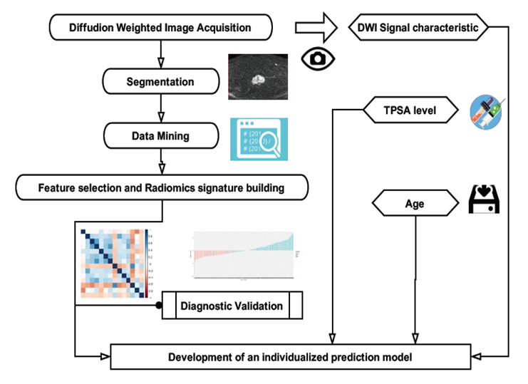
Figure 1.Workflow of the radiomics modeling
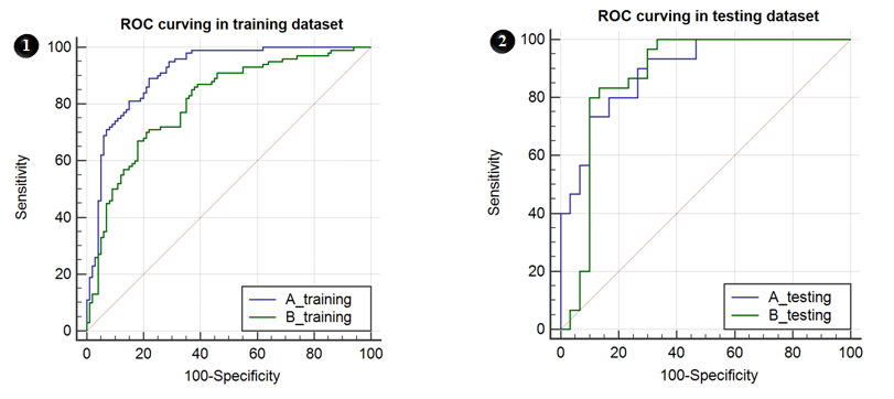
Figure 2. ROC curves in plan A&B, 6-1&6-2 were ROC
curves for training and testing dataset.
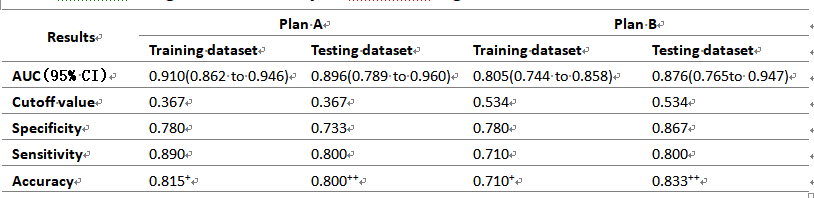
Table 1. Diagnostic efficiency
of Radiomic signature model in Plan A&B