3893
The change of amide proton transfer (APT) signal intensity (SI) with age in testes of adults: A preliminary study1Department of Radiology, the First Affiliated Hospital of Sun Yat-Sen University, Guangzhou, China, 2Philips Healthcare, Shanghai, China, 3Philips Healthcare, Guangzhou, China
Synopsis
Functional MRI, such as DWI and MTI, have been used to evaluate male infertility. Amide proton transfer (APT) is a novel MRI technique in the level of molecular imaging, which employs the chemical exchange of endogenous amide proton. This study investigated the relationship between amide proton transfer (APT) signal intensity (SI) changes with age in adults. According to the initial results of this research, we found that APT SI of testes was positively correlated age, which suggested that APT could be a potential biomarker for spermatogenic function.
Introduciton
Spermatogenic function evaluation is required in patients suspected of infertility. MRI allows noninvasive assessment of testicular lesions with high soft-tissue resolution and no ionizing radiation. Functional MRI, such as DWI and MTI, have been used to evaluate male infertility1-2. Amide proton transfer (APT) is a novel MRI technique in the level of molecular imaging, which employs the chemical exchange of endogenous amide proton. A previous study has revealed a negative relationship between APT values and age in brain development4. The aim of this study was to investigate the relationship between amide proton transfer (APT) signal intensity (SI) changes with age in adults.Materials and Methods
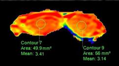
This study was approved by local ethics committee and written informed consent was obtained from each participant. Eleven healthy volunteers were enrolled and underwent MR scans on a 3.0T clinical scanner (Ingenia CX, Philips, Best, Netherlands) with a 32-channel Torso coil for signal reception. APT was performed with a three dimensional TSE-mDixon sequence with the main parameters as follows: saturation power=2µT , saturation duration=2s, frequency offsets= ±3.5, ±3.42, ±3.58, −1540 ppm, TR/TE/=6491/8.8ms, FOV=250×346mm2, voxelsize=1.8×1.8×4mm3, SENSE factor=1.6, scan duration=4:58min The APT weighted images were reconstructed online with intrinsic B0 maps after the acquisition was completed (Figure 1). Three ROIs were manually placed on the parametric maps of left and right testis respectively, and the mean values of each testis and of both testes were extracted. For the statistical analysis, data normality was determined by use of the Shapiro-Wilk test and then Pearson's or Spearman's correlations were used, as applicable. Statistical significance was defined as a p- value <0.05.Results
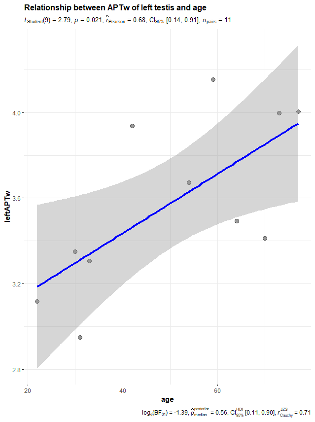
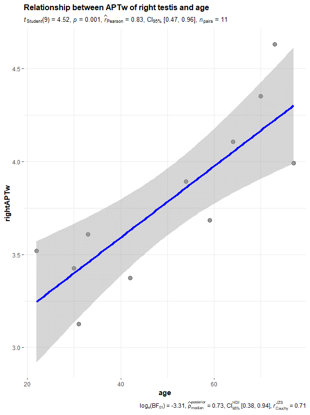
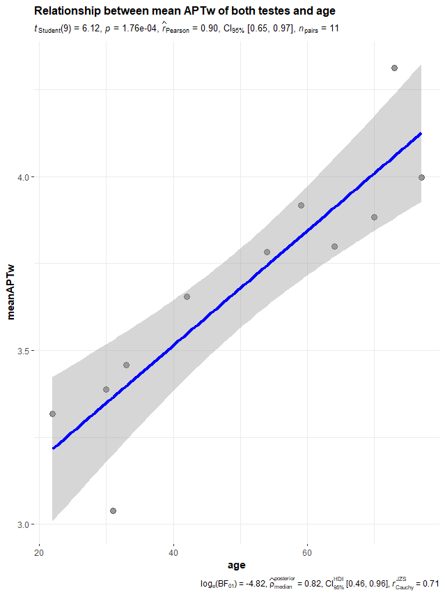
The mean age in the control group was 37 years (range: 22–59). The average APT singal intensity (SI) ± standard deviation (%) were 3.49±0.43 and 3.67±0.49 on the left-side and right-side testis, respectively. For the correlation analysis, we found that age was significant positive related to the APT SI both on the left testis (r=0.68, p=0.021) and the right testis (r=0.83, p=0.001), as shown in Fig.2 and Fig.3, respectively. Further, we also found that there was a positive correlation between the age and the mean APT SI of both testes (r=0.90, p < 0.001) (Fig.4).Discussion and Conclusions
As APT imaging is sensitive to the level of mobile proteins in biological tissues, the increase of APT SI with age may indicate the decreased level of solid protein for spermatogenesis. Although further study is required, the initial results of this research shows that APT SI of testes are positively correlated with age, and APT SI of testes could be a potential biomarker for spermatogenic function.Acknowledgements
No acknowledgement found.References
[1] Tsili AC, Ntorkou A, Baltogiannis D, et al. Magnetization transfer imaging of normal and abnormal testis: preliminary results. Eur Radiol, 2016;26:613–621.
[2]Wang H, Jian Guan, Lin JH, Guo Y, Li SR, Cai HS. Diffusion weighted and magnetization transfer imaging in testicular spermatogenic function evaluation: preliminary results. Journal of Magnetic Resonance Imaging. 2018,47(1):186-190.
[3] Zhou J, Payen J, Wilson D, Traystman R, van Zijl PC. Using the amide proton signals of intracellular proteins and peptides to detect pH effects in MRI. Nat Med, 2003, 9: 1085-1090.
[4] Zhang H, Kang H, Zhao X, Jiang S, Zhang Y, Zhou J, Peng Y. Amide Proton Transfer (APT) MR imaging and Magnetization Transfer (MT) MR imaging of pediatric brain development. Eur Radiol. 2016 Oct;26(10):3368-76.
[5] Wen Z, Hu S, Huang F,et al. MR imaging of high-grade brain tumors using endogenous protein and peptide-based contrast. Neuro-Image,2010, 51:616–622
Figures