3892
Inhibition of Urine Amide Proton Transfer Signal with Optimal Scan Parameters in Bladder Cancer1Henan Provincial People's Hospital, Zhengzhou, China, 2Philips healthcare, Beijing, China
Synopsis
In the preliminary experiment, we found that there were hyperintense amide proton transfer weighted (APTw) signal in urine, which may interfere with the contrast of APTw signal in bladder lesion. In this study, we aim to explore the feasibility and features of urine APTw imaging in vitro and in vivo, and inhibite the urine APTw signal for better display of bladder lesions using optimal parameters.
Synopsis
In the preliminary experiment, we found that there were hyperintense amide proton transfer weighted (APTw) signal in urine, which may interfere with the contrast of APTw signal in bladder lesion. In this study, we aim to explore the feasibility and features of urine APTw imaging in vitro and in vivo, and inhibite the urine APTw signal for better display of bladder lesions using optimal parameters.Introduction
Bladder cancer is the most common malignant tumor of urinary system. In clinical practice, it is difficult to differentiate benign from malignant bladder cancer, especially in early phase. In the preliminary experiment, we found that there were hyperintense amide proton transfer weighted (APTw) signal in urine, which may interfere with the contrast of APTw signal in bladder lesion. In this study, we aim to explore the feasibility and features of urine APTw imaging, and inhibite the urine APTw signal for better display of bladder lesions using optimal parameters.Materials and Methods
Four tube contain with urea of the concentration of 2 %, 4 %, 6 %, 8 % and two tubes with distilled water and egg white as the baseline were used to test the feasibility of urea APT imaging. Other two tubes contain urine from a healthy volunteer and a patient with bladder cancer were detected to prove the possible difference. These tubes were fixed in agarose (3%) to reduce the interference to B0 from air around. 2 male patients with bladder lesion were recruited. APTw imaging were performed on a 3.0 T MR scanner (Ingenia CX, Philips Healthcare, the Netherlands). In order to reduce the signal of utine APTw, the optimal scan parameters (including saturated B1 power and duration time, voxel and SENSE) were established. Raw data of APTw were uploaded to a workstation (Intellispace Portal 9, Philips Healthcare) for post-processing.Results
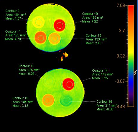
In phantom, the averaged signal intensity of APTw with urea of the concentration of 2 %, 4 %, 6 %, 8 % were 1.07%, 2.46%, 4.78%, 7.22%, respectively. The APTw signals in water and egg white were -0.38% and 8.25%. In human urine in vitro, the APTw signals of were a healthy volunteer and a patient with bladder cancer were 3.13% and 0.29%, respectively. As for patients with bladder lesion, the better APTw contrast in bladder lesions, lower image distortion and artifacts was observed with B1 of 1.5ut, B1 duration of 1.5s and voxel of 3×3 mm2.Discussion and conclusion
In this preliminary study, we found that APTw signal increased with elevated concentrations of urea in phantoms, suggesting the hyperintense amide proton transfer weighted (APTw) signal in urine may associated with the protein and urea in urine. Abnormal low APTw signal intensity in urine of a patient with bladder cancer in vitro may attribute to its residence for a longer time, prompting the fresh urine is necessary of the in vitro experiment. With the optimal scan parameters, the higher APTw contrast in bladder lesions was acquired, which is helpful to differentiate benign from malignant bladder cancer using APTw imaging.Acknowledgements
No acknowledgement found.References
1.Su C , Liu C , Zhao L , et al. Amide Proton Transfer Imaging Allows Detection of Glioma Grades and Tumor Proliferation: Comparison with Ki-67 Expression and Proton MR Spectroscopy Imaging[J]. Ajnr American Journal of Neuroradiology, 2017.
2.Nishie A , Takayama Y , Asayama Y , et al. Amide proton transfer imaging can predict tumor grade in rectal cancer[J]. Magnetic Resonance Imaging, 2018, 51:96-103.
3.Keisuke, Ishimatsu, Akihiro, et al. Amide proton transfer imaging for differentiating benign ovarian cystic lesions: Potential of first time right[J]. European journal of radiology, 2019, 120:108656-108656.
4.Li B , Sun H , Zhang S , et al. Amide proton transfer imaging to evaluate the grading of squamous cell carcinoma of the cervix: A comparative study using 18F FDG PET[J]. Journal of Magnetic Resonance Imaging, 2019, 50(1).
5.A B L , A H S , A S Z , et al. The utility of APT and IVIM in the diagnosis and differentiation of squamous cell carcinoma of the cervix: A pilot study[J]. Magnetic Resonance Imaging, 2019, 63:105-113.
Figures