3884
Use of intravoxel incoherent motion MRI to assess placental perfusion in normal and Fetal Growth Restricted pregnancies on their third trimester
liu xilong1, Feng jie1, Huang chantao1, Mei Yingjie2, and xu yikai1
1Department of Medical Imaging Center, Nanfang Hospital, Southern Medical University, Guang Zhou, China, 2Clinical science, Philips Healthcare, Guangzhou, China
1Department of Medical Imaging Center, Nanfang Hospital, Southern Medical University, Guang Zhou, China, 2Clinical science, Philips Healthcare, Guangzhou, China
Synopsis
Early diagnosis of FGR is vitally important for the management of pregnancy and delivery planned accordingly. IVIM MRI is a non-invasive, in vivo techniques which can assess placental perfusion quantitatively, and be useful for evaluating placental microcirculation. In this study, the IVIM parameters of placental was measured and to investigate whether FGR pregnancies have different placental perfusion compared with normal pergnancies. The results show that perfusion fraction (f) have a significantly lower in FGR group. And f value moderately incresased with increasing gestational age in normal pregnancies.
Purpose and introuduction
Diagnosis and monitoring of Fetal Growth Restricted (FGR) is currently limited to measuring fetal biometry and heart rate, amniotic fluid volume, and assessment of blood flow using Doppler ultrasound. And at the point of diagnosis with ultrasound there has already been damage to placental microstructure and function1,2. Therefore, it is important to identify FGR as early as possible, so that the fetus can be closely monitored and the delivery planned accordingly. Intravoxel incoherent motion (IVIM) magnetic resonance imaging is a novel method which can assess placental perfusion quantitatively2,3. Therefore, the first aim of this study was to investigate whether FGR pregnancies have different placental perfusion compared with normal pergnancies. A secondary aim was to investigate if the microvascular perfusion fraction of the placenta changes with increasing gestational age in normal pregnancy.Materials and Methods
The study population included 17 FGR pregnancies (average age, 29 years; range, 19-38 years) and 36 normal pregnancise (average age, 29.2 years; range, 16-40 years) between 28+3 to 38+0 weeks. All women underwent a MRI examination including an IVIM sequence at 3.0 T MRI system (Ingenia 3.0 T; Philips Health care, Best, the Netherlands). IVIM sequence was used acquisition matrix 304×230, FOV 400×364 mm, slice thickness 5 mm, number of slices 36, and 9 different b-values (0,10,20,40,80,200,400,600,1000 s/m2) perpendicular to the placenta. The total acquistion time was 9 min 30 s. Evaluation of the IVIM sequence was performed with research software (AW_MADC). ROIs were placed in the middle part of the placenta including as large parts of the placenta as possible, but excluding areas with infarcts, hemorrhage or other artifactual signal loss (Figure 1). The same ROIs were draw on the slice above and below the middle slice. We calculated the values of the standard diffusion coefficeint (D), pseudodiffusion (D*) and perfusion fraction (f) by averaging over 3 ROIs totally. Student t test and chi-square were used for statistical analysis. Spearman correlation coefficient was calculated between f and gestational age in normal pregnancy. P < 0.05 was considered to be a statistically significant difference.Results
The perfusion fraction (f) was significantly lower in the FGR group than that in the normal group (33.96±2.62(%) vs 38.48±5.31(%), p = 0.002). Differences of standard diffusion coefficeint (D) and pseudodiffusion (D*) in two groups showed no statistical significance (P > 0.05). The placental perfusion fraction (f) moderately incresased with increasing gestational age in normal pregnancies (R2 = 0.173, p = 0.01) (Figure 2).Conclusion
The perfusion fraction (f) is able to distinguish FGR from normal pregnancies. It can be used as a feasible index to evaluate placenta perfusion. Placental perfusion moderately increases with incerasing gestational age in normal pregnancy.Acknowledgements
No acknowledgements found.References
- Chen T, Zhao M, Song J, et al. The effect of maternal hyperoxygenation on placental perfusion in normal and Fetal Growth Restricted pregnancies using Intravoxel Incoherent Motion. Placenta. 2019 Dec;88:28-35. doi: 10.1016/j.placenta.2019.08.078.
- Slator PJ, Hutter J, McCabe L, et al. Placenta microstructure and microcirculation imaging with diffusion MRI. Magn Reson Med. 2018 Aug;80(2):756-766. doi: 10.1002/mrm.27036.
- Jakab A, Tuura RL, Kottke R, et al. Microvascular perfusion of the placenta, developing fetal liver, and lungs assessed with intravoxel incoherent motion imaging. J Magn Reson Imaging. 2018 Jul;48(1):214-225. doi: 10.1002/jmri.25933.
Figures
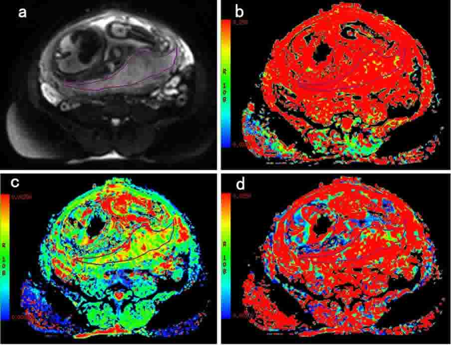
An example of IVIM
result from a FGR pregnancy (38 years, 31 gestational weeks). a) diffusion
weighted image at b-value = 0 mm2/s; b) perfusion fraction (f)
map, with f value of 36.4%; c) standard diffusion coefficeint (D) map, with
D value of 1.46×10⁻³mm2/s; d) pseudodiffusivity (D*) map, with D* value of 19.9×10⁻³mm2/s
. The ROI was draw on diffusion weighted image (b = 0 mm2/s) and
then transferred to the other three maps.
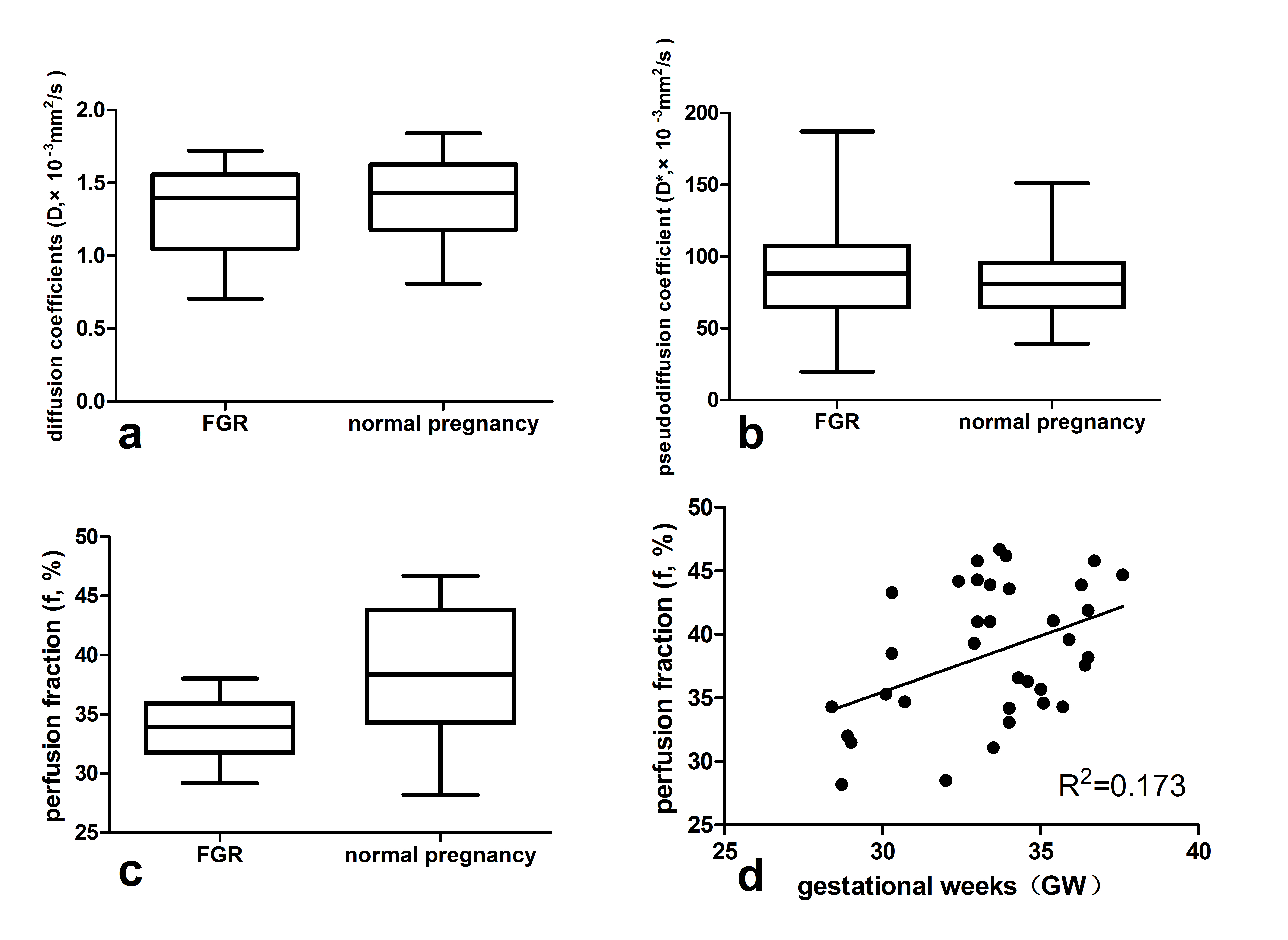
a-c. Box plot of IVIM
parameters for normal and FGR groups. a) standard diffusion coefficeint (D);
b). pseudodiffusivity (D*); c) perfusion fraction (f). d) Scatter plot showing
the perfusion fraction (f) in relation to gestational age in normal pregnancy
(p = 0.01). The estimated regression line is shown in the scatter plot (R2
= 0.173).