3881
Correlation between Ki67 expression and Intraventricular incoherent motion (IVIM) in ovarian cancer
Xinliu He1, Yulin Chen1, Li Liu1, Ye Li1, Qingwei Song1, and Ailian Liu1
1The First Affiliated Hospital of Dalian Medical University, Dalian, China
1The First Affiliated Hospital of Dalian Medical University, Dalian, China
Synopsis
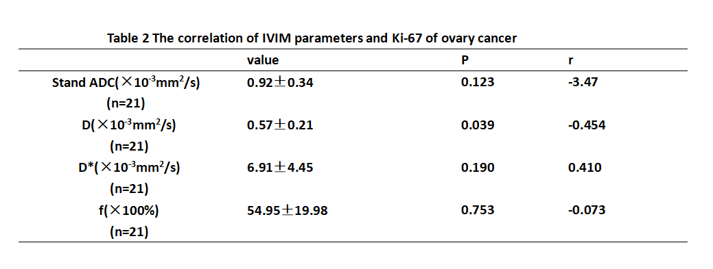
Ki-67 is one of the prognostic marker which determines the growth fraction of a tumour and its over expression is associated with malignancy, tumour aggression, reserved prognosis and metastasis. This worked aimed at exploring the correlation of Intravoxel incoherent motion (IVIM) quantitative parameters with ki-67 expression level in ovarian cancer. The results proved that there are correlation between D value and Ki-67 gene expression in ovarian cancer, P value is 0.039 and the r value is -0.454.
Introduction
Ovarian cancer (OC) is one of the most common tumors in the female reproductive system. The incidence rate of ovarian cancer ranks third in the female reproductive system malignant tumors, and the mortality rate ranks first. Proliferating nuclear antigen Ki67 reflects the proliferative activity of tumors and is widely used to predict the prognosis of many tumors. IVIM can be used to evaluate the pure molecular diffusivity (performed by the parameters apparent diffusion coefficient [ADC] and diffusion coefficient value [D]) and perfusion-related diffusivity (performed by parameters perfusion coefficient value [D*] and perfusion fraction value [f]). Therefore, the purpose of this study is to evaluate the efficacy of IVIM parameters in the preoperative evaluation of ki-67 expression in OC.Methods
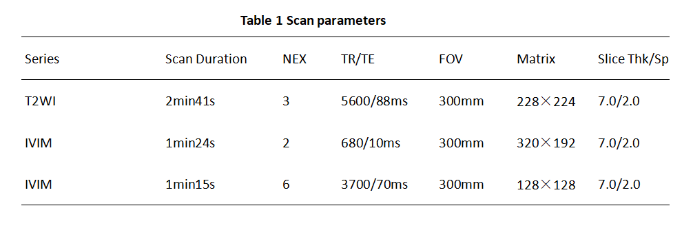
A total of 20 patients (age: 54.75 ± 12.99 years range (24-76 years) )with 21 lesions of ovarian cancer confirmed by surgery and pathology from 2015 to 2020 were retrospectively collected. This research Includes 10 serous cystadenocarcinoma with 11 lesions, 1 mucinous cystadenocarcinoma, 2 endometrioid adenocarcinoma, 3 clear cell carcinoma, 1 metastasis, 1 endometrial stromal sarcoma, 1 mixed germ cell tumor, and 1 adult granulosa cell tumor patients. All patients underwent abdominal MR examinations (Signa HDxt, GE Medical Systems, USA) included T2WI, IVIM and LAVA(Scan parameters show in table 1). The original axial digital images from the IVIM sequence were transmitted to the GE SDC-ADW 4.6 workstation (Sun Microsystems, Santa Clara, Calif) and the post-processing was performed by Functool software. Referring to the anatomical location of lesion obtained on T2-weighted or DWI images, The stand ADC, D, D* and f maps were automatically constructed, and were reviewed by two observers who were blinded to clinical information and histopathologic results with 10 and 15 years of experience in pelvic imaging, respectively. The manual regions of interest (ROIs) were drawn along the edge of tumors on the slice with maximal solid area (we choose the solid in tumors), according to fat suppression T2WI and T1WI (Fig. 1). The measurement was repeated for three times, and the averages of three measurements were calculated. The stand ADC, D, D* and f were recorded. Correlation between above values and the expression of Ki-67 in gastric cancer was analyzed by Pearson correlation coefficient test..Results
ICC for all parameters were more than 0.75(table2). The Ki-67 expressions in cancer tissue was negatively correlated with cancer D values (r=-0.454, P=0.039. while Ki-67 expression was not correlated with stand ADC, D* and f value of ovary cancer. (table2)Discussion and Conclusion
Many studies have investigated immunohistochemical expression of Ki-67 as a prognostic and predictive marker for cancer [1-3]. The D value reflects the diffusion state of water molecules in tissues. Because the degree of tumor differentiation is related to cell heteromorphism, the lower the degree of differentiation, the greater the cell heteromorphism; the active proliferation of tumor cells leads to the increase of microvessel density, which leads to the limitation of water molecule diffusion. So, our research show D value has a negative correlation with Ki67 value. The D value of IVIM can predict Ki-67 status of ovarian cancer.Acknowledgements
No acknowledgement found.References
1.Alexey Surov,Hans Jonas Meyer,Andreas Wienke.Associations between apparent diffusion coefficient (ADC) and KI 67 in different tumors: a meta-analysis. Part 1: ADCmean.[J].Oncotarget,2017,8(43).75434-75444.2.Xiaoyu Zeng,Lin Liu,Mengzhu Zheng,et al.Pantoprazole, an FDA-approved proton-pump inhibitor, suppresses colorectal cancer growth by targeting T-cell-originated protein kinase.[J].Oncotarget,2016.22460-73.3.Feng Wang,Yuxiang Wang,Yan Zhou,et al.Apparent Diffusion Coefficient Histogram Analysis for Assessing Tumor Staging and Detection of Lymph Node Metastasis in Epithelial Ovarian Cancer: Correlation with p53 and Ki-67 Expression[J].Molecular Imaging & Biology,2019,21(4).731-739.Figures
.
.
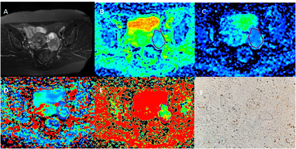
Figure 1 A 47-year-old woman with left side ovarian cancer. A T2WI shows a low signal lesion in left pelvis .B The stand ADC value is 0.713×10⁻³mm2/s. C The D value is 0.488×10⁻³mm2/s. D The D* value is 10.7×10⁻³mm2/s. E The f value is 0.371. F The Ki-67 picture of this lesion.