3866
Robust Fat Suppression with Slice Selective Gradient Reversal (SSGR) in Amide Proton Transfer Imaging – Application to Non-Human Primate Brain1Yerkes Imaging Center, Yerkes National Primate Research Center, Emory University, Atlanta, GA, United States, 2Department of Radiology and Imaging Sciences, Emory University School of Medicine, Atlanta, GA, United States, 3Center for Visual and Neurocognitive Rehabilitation, Atlanta VA, Decatur, GA, United States, 4Department of Physics & Astronomy, Georgia State University, Atlanta, GA, United States, 5Department of Radiology, University of Alabama Birmingham, Birmingham, AL, United States
Synopsis
Single-shot EPI imaging has become a widely used acquisition method in amide proton transfer (APT) imaging due to its fast imaging readout. Because APT effects are relatively small, fat artifacts may significantly impact the asymmetry analysis. Recently, a slice selective gradient reversal (SSGR) method has been reported to effectively suppress fat signals. This work aims to introduce the SSGR method into spin-echo EPI APT imaging, which can be used for fat suppression in the non-human primate brain with substantial pericranial fat.
Introduction
Single-shot echo planar Imaging (EPI) has become a widely used acquisition method in amide proton transfer (APT) imaging due to its fast imaging readout.1 However, its low bandwidth in the phase-encoding direction makes it susceptible to chemical-shift artifacts from lipids. Because APT effects are relatively small, fat artifacts may significantly impact the asymmetry analysis. Although most APT images are acquired with a chemical shift selective fat saturation technique to suppress unnecessary fat signal, the remains of the lipid signal can still be observed on some image slices. Recently, a slice selective gradient reversal (SSGR) method has been reported to effectively suppress fat signals.2,3 This work aims to introduce the SSGR method into spin-echo EPI APT imaging, which can be used for fat suppression in the non-human primate brain with substantial pericranial fat.Theory
The SSGR method can be implemented for 2D multislice APT imaging as illustrated by the configuration in Fig.1a, where the reversed gradient (red) is used in the refocusing radio frequency (RF) pulse instead of a normal slice selective gradient (red dotted line). The displacement of the excited fat slice relative to the water slice (Fig.1b) is$$\tt D=\frac{\tt \delta \cdot \tt B_{\tt 0}}{\tt G_{\tt z}}$$
where $$$\tt \delta$$$ is the chemical shift (ppm), $$$\tt B_{\tt 0}$$$ is the static magnetic field (T), and $$$\tt G_{\tt z}$$$ is the slice-selective gradient amplitude (T/m). Because the slice-selective gradient amplitude is proportional to the RF bandwidth (BW), the displacement can be controlled by changing the RF bandwidth while maintaining the slice thickness.
Methods
We collected images from a creatine phantom and non-human primate brain with a 3T Siemens whole-body MAGNETOM Trio scanner (Siemens Medical Solution). 50mM creatine and oil were transferred to falcon tube, respectively. The phantom holder was filled with 1.5% agarose gel. The common MRI parameters of the proposed method for the phantom study and the non-human primate scan were: B1 = 0.7 uT, offset frequencies from -5 to 5 ppm with increments of 0.1 ppm, Tsat/Trec = 2/2 sec, duration of EPI module is 38 ms, and the base resolution is 2 x 2 x 5 mm3. In the conventional SSGR method, excitation RF BW (BWex) and refocusing RF BW (BWrf) were originally set to 850 and 600 Hz, respectively. The redesigned SSGR method used BWex/BWrf = 425/300 Hz by considering appropriate displacement between water and fat. For the phantom study, five sets of APT imaging were obtained as follows: 1) with fat suppression, 2) with SSGR, 3) with both fat suppression and SSGR, 4) with redesigned SSGR based on the low RF bandwidth, and 5) with both fat suppression and redesigned SSGR. Total imaging time is 13 min per set with a single slice and 2 averages. Three sets of the non-human primate brain imaging, including 1) using fat suppression, 2) using both fat suppression and SSGR, and 3) using both fat suppression and redesigned SSGR. Total imaging time took 28 min per set with six slices and 4 averages.Results/Discussion
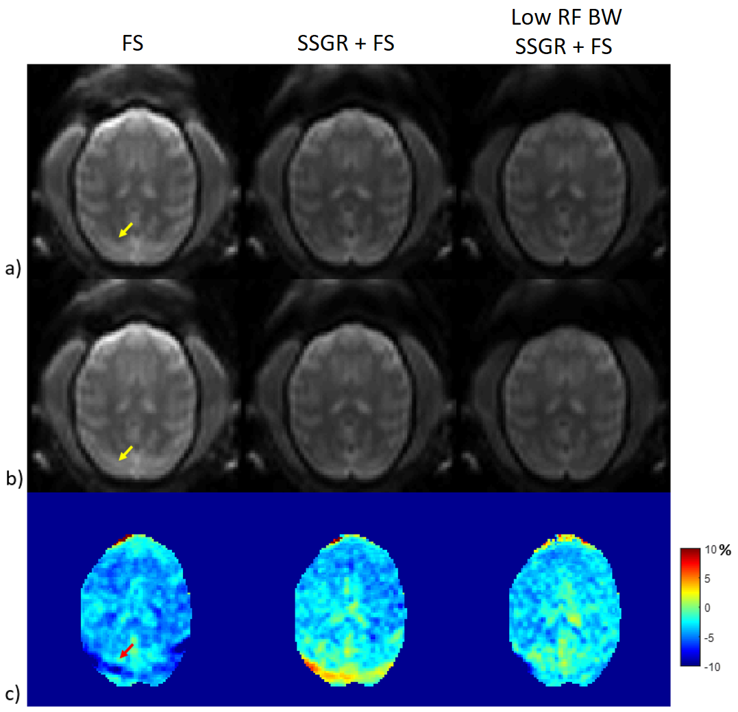
Figs.2a-c show magnitude images of the creatine phantom at 0, -1.9, and 1.9 ppm , respectively. It is noted that the remains of the fat signal are visible when using both fat suppression and SSGR designed from inappropriate RF bandwidth (Fig. 2c). Such fat artifacts have a significant impact on the asymmetric analysis (Fig.2d). Fig.3 shows APT images of non-human primate brains. As compared with the conventional methods, the proposed method can remove fat signals from the pericranial area. It is noted that an additional T2* effect may occur during the proposed method acquisition because the echo time was increased by 2ms due to the RF duration elongation.Conclusion
We demonstrated the effect of the SSGR method on fat suppression in APT imaging with a phantom and non-human primate study. It is expected that the proposed method can be further extended to clinical applications including muscle, knee, and breast imaging.Acknowledgements
No acknowledgement found.References
[1] Zhou J, et al. Using the amide proton signals of intracellular proteins and peptides to detect pH effects in MRI, Nat Med 2003;9:1085-90.
[2] Zoltan N, et al. Efficient fat suppression by slice-selection gradient reversal in twice-refocused diffusion encoding. MRM 2008 60:1256-1260
[3] Takahara T, et al. Fat suppression with slice-selection gradient reversal (SSGR) revisited, ISMRM 2009
Figures
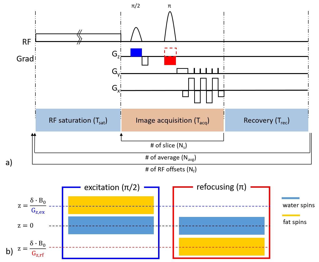
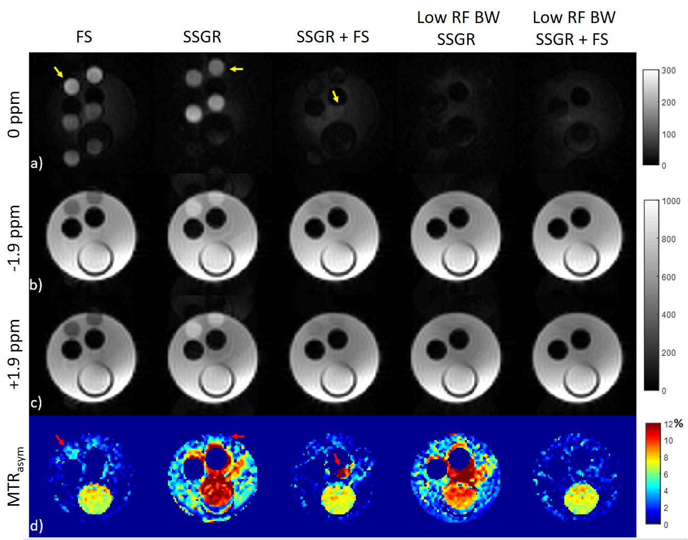
Fig 2. Comparison of the creatine-fat phantom images acquired using fat suppression (first column), SSGR based on BWex/BWrf = 850/600 Hz (second column), both fat suppression and SSGR (third column), redesigned SSGR based on BWex/BWrf = 425/300 Hz (fourth column), and both fat suppression and resedigned SSGR (fifth column). Magnitude images at a) 0 ppm, b) -1.9 ppm, c) 1.9 ppm. d) The corresponding MTR asymmetry map.