3844
Three-Point Dixon Abdominal Water/Fat Separation using a Lower-Field 0.75T MRI1University and ETH Zurich, Zurich, Switzerland, 2Philips Research, Hamburg, Germany, 3Leiden University Medical Center, Leiden, Netherlands
Synopsis
Water/fat separation can be performed by exploiting chemical shift, which is proportional to field strength. Hence, on lower-field MRI systems (0.1 … 1T), the absolute resonance frequency difference is reduced compared to clinical high-field MRI, raising the question if water/fat separation can still be reliably performed despite the proximity of the resonances. In this work, we examined the feasibility of Dixon-type water/fat separation in the abdomen on a 0.75T lower-field MRI system. We show that Cartesian multi-acquisition and multi-echo as well as spiral multi-acquisition sequences provide reliable water/fat separation despite the reduced water/fat resonance shift of approximately 108 Hz.
Introduction
Dixon water/fat separation relies on the chemical shift of protons in water and fat.1 With the recent advent of lower-field MRIs (0.1…1T), the question arises if the proximity of the water and fat resonance frequencies (108 Hz at 0.75T) allows for reliable water/fat separation. In this work, we evaluate the feasibility of abdominal chemical shift-based water/fat separation on a 0.75T lower-field MRI using three-point Dixon with Cartesian and spiral readoutsMethods
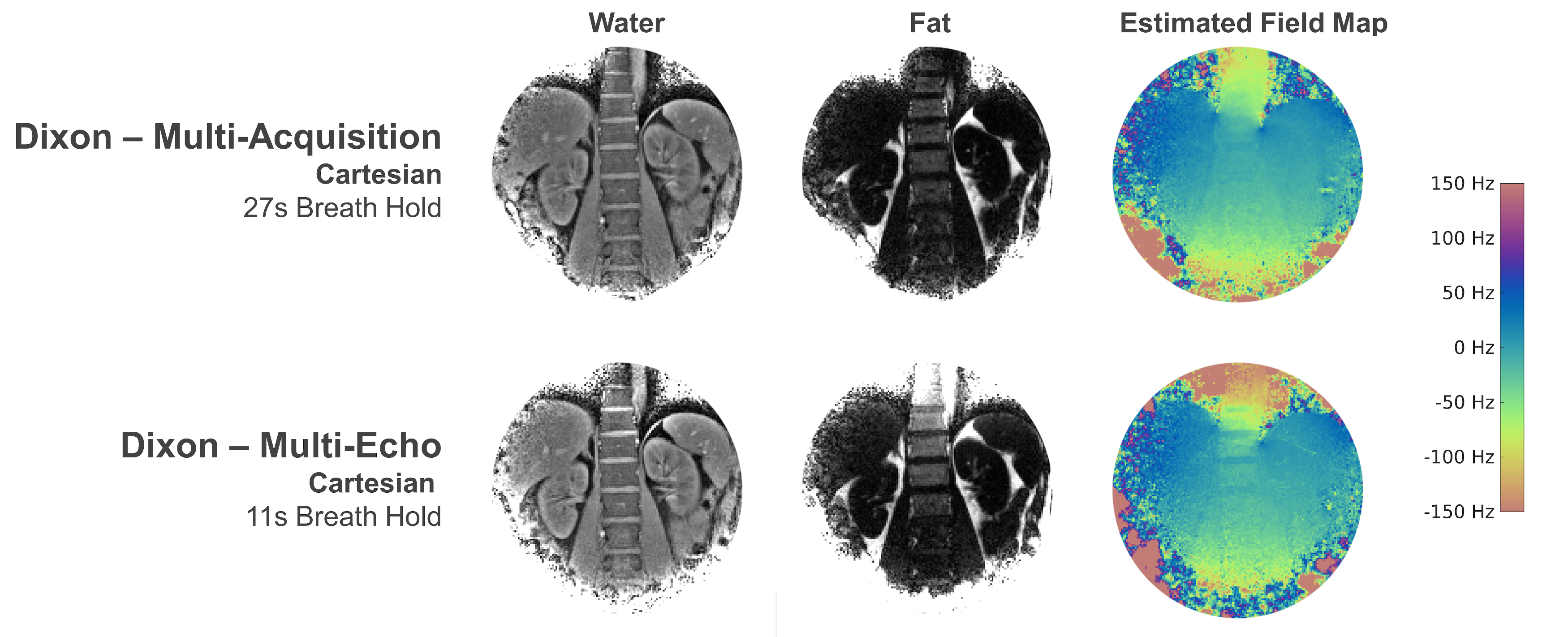
We exploited the Carbon-13 channels of the multi-nuclear interface of a 3T Philips Achieva system (Best, The Netherlands) to measure proton magnetization at a quarter of the nominal field strength when the system was ramped to 0.75T. A Helmholtz-like volume transmitter and a four-channel receive array designed to support 13C at 3T (Clinical MR Solutions, Brookfield, WI, USA) was used for excitation and signal reception.Cartesian multi-acquisition (MA) and multi-echo (ME) Dixon scans were performed in healthy volunteers by adapting the echo time increments of the Philips product sequence to 0.75T. The first echo time was set to 2.9ms for MA (4ms ME) with an echo time increment of 3.07ms in accordance with 3-point Dixon methods2,3 and an average chemical-shift of water and fat of 108 Hz at 0.75T. Additional scan parameters were: single slice in coronal orientation; 176x176 matrix size; field-of-view (FOV): 350mm; slice thickness: 8mm; TR: 11.5ms (MA), 12.1ms (ME); FA: 15°; Number of Averages: 4. The resulting nominal resolution for the Cartesian scans was 2mm in-plane.
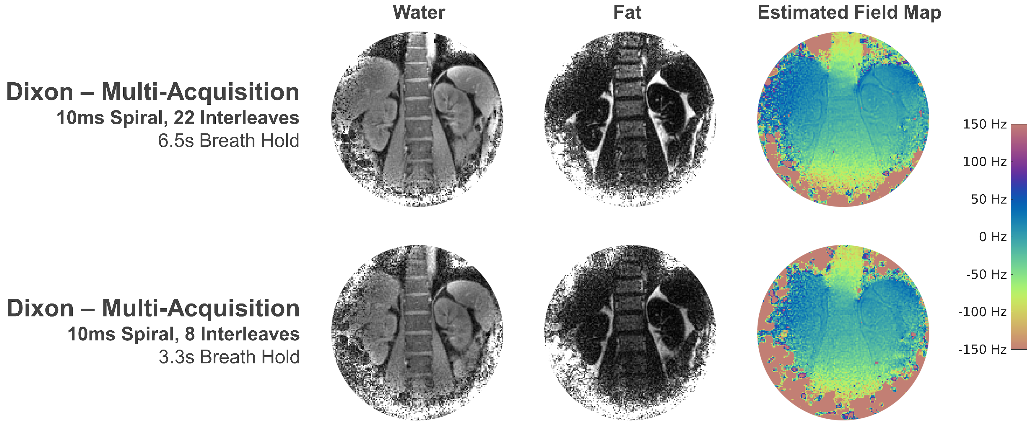
In addition, spiral MA Dixon scans were performed using 10ms Archimedean spirals with 8 and 22 interleaves leading to 4- and 11-fold averaging, respectively. The echo time was set to 1.1ms resulting in a TR of 18.8ms and otherwise comparable scan parameters as the Cartesian MA scan.
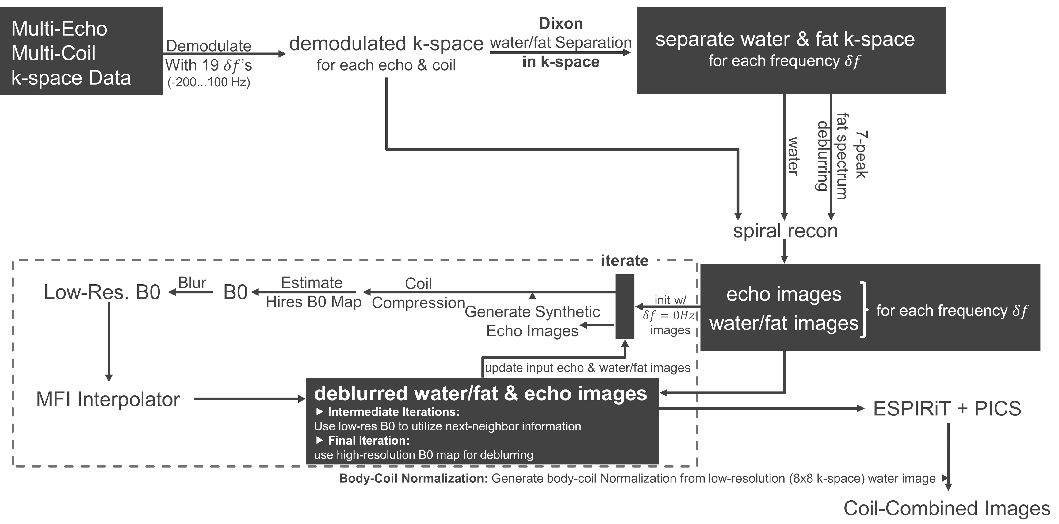
Reconstruction of the spiral data was performed in MATLAB 2018a (Mathworks, Natick, MA, USA), MRecon (GyroTools LLC, Winterthur, Switzerland), and BART4 using an iterative scheme as shown in Figure 1. Water/fat separation was performed in k-space and the fat-channel was deblurred using a seven-peak fat-spectrum.5,6 Reconstructed echo and water/fat images were fed into an iterative reconstruction pipeline to estimate a B0 map by comparing the phase of forward simulated and acquired echo images; subsequently, B0 deblurring using multi-frequency interpolation was performed7 including feedback of the deblurred echo and water/fat images into the B0 estimation code. Coil combination was performed using ESPIRiT8 and Bart’s PICS code. Due to the lack of a uniform body coil receive pattern, homogenization was performed by dividing by a low-resolution (8x8 k-space) reconstruction of the water channel.
Cartesian reconstruction was performed equivalently, however, omitting the spiral reconstruction steps as well as B0 deblurring. Here, fat-spectrum deblurring was performed directly in image space as part of the water/fat separation step involving the fat seven-peak model.
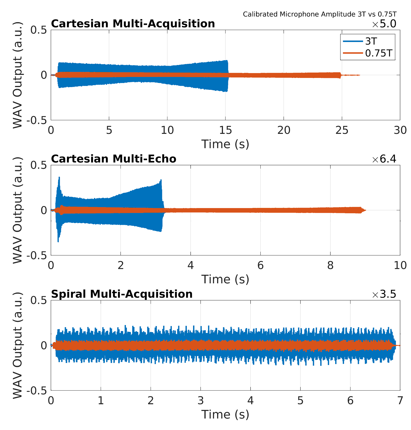
Apart from imaging experiments, audio measurements in the scanner room were performed to compare sound pressure levels between lower-field and product field-strength on the same scanner. The microphone (Sennheiser ME 66) and linear recorder (Tascam DR-100) were calibrated using a 1kHz tone. For comparison to 3T, the time of the Dixon sequences was adapted to the higher field strength leading to an echo time shift of 0.77ms and an according reduction of TR.
Results & Discussion
Breath hold duration for a single slice was 27s for the Cartesian MA and 11s for the ME scan, respectively. Good water/fat separation performance was achieved using both techniques and comparable field maps were estimated (Figure 2).Breath hold durations could be further reduced by employing spiral readouts in the multi-acquisition Dixon scan, resulting in scan durations of 6.5s and 3.3s, respectively. Again, chemical shift-based separation of water and fat was achieved as well as blurring-free spiral water and fat images (Figure 3).
Due to the limited receive field of the four-channel cardiac receive array, noise amplification is observed at the boundary of the field-of-view. Also, transmit-field inhomogeneity can be observed (e.g. hyper-intensity at the spleen), which is due to the inhomogeneous B1+ of the employed transmitter. The former can be overcome by employing abdominal arrays with increased coverage, while transmit field homogeneity can be considerably improved by using a body coil instead of the flexible Helmholtz-like two-loop transmitter.
In Figure 4, time vs. microphone output plots are shown for the three sequence types, yielding a 3- to 6-fold higher amplitude on 3T relative to 0.75T. Especially in the 3T multi-echo case, the echo spacing of 0.77 ms is close to a larger mechanical resonance of the Philips Achieva gradient system (1.2 kHz) yielding elevated sound pressure levels. The reduction of Lorentz forces acting on the gradient system and generally more lenient sequence characteristics of the lower-field system lead to intrinsically lower sound pressure levels.
Conclusions
We have demonstrated feasibility of chemical-shift based water/fat separation using both multi-acquisition and multi-echo Dixon with Cartesian and spiral readouts at the reduced field strength of 0.75T within clinically feasible breath hold durations. We could further demonstrate that Dixon imaging benefits greatly from the reduced field strength in terms of reduced sound pressure level, which increases patient comfort. Future work will be focused on volumetric imaging and free breathing acquisitions.Acknowledgements
No acknowledgement found.References
1. Dixon WT. Simple proton spectroscopic imaging. Radiology. 1984;153(1):189-194. doi:10.1148/radiology.153.1.6089263
2. Glover GH, Schneider E. Three-point dixon technique for true water/fat decomposition withB0 inhomogeneity correction. Magn Reson Med. 1991;18(2):371-383. doi:10.1002/mrm.1910180211
3. Glover GH. Multipoint dixon technique for water and fat proton and susceptibility imaging. J Magn Reson Imaging. 1991;1(5):521-530. doi:10.1002/jmri.1880010504
4. BART Toolbox for Computational Magnetic Resonance Imaging. doi:10.5281/zenodo.592960
5. Brodsky EK, Holmes JH, Yu H, Reeder SB. Generalized K-space decomposition with chemical shift correction for non-Cartesian water-fat imaging. Magn Reson Med. 2008;59(5):1151-1164. doi:10.1002/mrm.21580
6. Yu H, Shimakawa A, McKenzie CA, Brodsky E, Brittain JH, Reeder SB. Multiecho water-fat separation and simultaneous R 2* estimation with multifrequency fat spectrum modeling. Magn Reson Med. 2008;60(5):1122-1134. doi:10.1002/mrm.21737
7. Man L-C, Pauly JM, Macovski A. Multifrequency interpolation for fast off-resonance correction. Magn Reson Med. 1997;37(5):785-792. doi:10.1002/mrm.1910370523
8. Uecker M, Lai P, Murphy MJ, et al. ESPIRiT - An eigenvalue approach to autocalibrating parallel MRI: Where SENSE meets GRAPPA. Magn Reson Med. 2014;71(3):990-1001. doi:10.1002/mrm.24751
Figures