3821
High-Performance Low-Field MRI of the Lumbar Spine: Comparison of 0.55T MRI With Two Gradient Systems To 1.5T MRI in Humans1Department of Radiology, NYU Langone Medical Center, New York, NY, United States, 2Siemens Medical Solutions USA Inc, Malvern, PA, United States
Synopsis
Next-generation low-field magnetic resonance imaging (MRI) systems hold great potential to reduce the cost of ownership and improve access to MRI worldwide. We compared MRI of the lumbar spine at 0.55T MRI with two different gradient performance modes to 1.5T MRI in 10 volunteer participants. Regardless of field strength and gradient performances, all MRI studies had sufficient signal-to-noise-ratios, contrast-to-noise-ratios, image quality, and visibility of anatomical structures with only small differences. Our initial results support the great potential of next-generation low-field MR systems to achieve similar image quality than state-of-the-art clinical 1.5-T MRI systems to detect the lumbar spine abnormalities.
Introduction
Next-generation low-field magnetic resonance imaging (MRI) systems can be built more powerful and cost-effective, holding great potential to reduce the cost of ownership and improve access to MRI worldwide.1,2 State-of-the-art magnet technology with 0.55T field strength, radiofrequency coils, gradient systems, pulse sequence techniques, receiver coils, signal conduction and conversion, and processing afford new possibilities to narrow the gap to clinical 1.5T MRI systems. The advantages of an advanced low-field 0.55-T MRI system have recently been demonstrated for interventional MRI2. While the inherently shorter T1 tissue constants and lower magnetic susceptibility effects are favorable for spine MRI, the remaining lower signal-to-noise ratios and naturally prolonged T2 constants represent formidable challenges. Few studies comparing the diagnostic capability of 0.2T low-field MRI for lumbar degenerative disease described challenges in achieving appropriate image quality, cerebrospinal fluid brightness, and clinically viable acquisition times scan3–5. The purpose of our study was to test the hypothesis that a high-performance, low-field-strength 0.55-T MRI system can generate MR images of the lumbar spine with similarly sufficient image quality and visualization of anatomical structures than state-of-the-art clinical 1.5T MRI.Methods
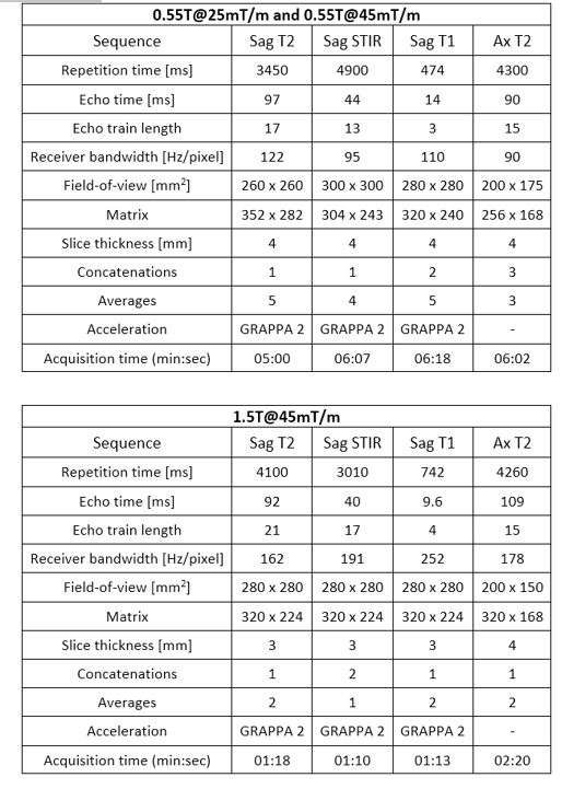
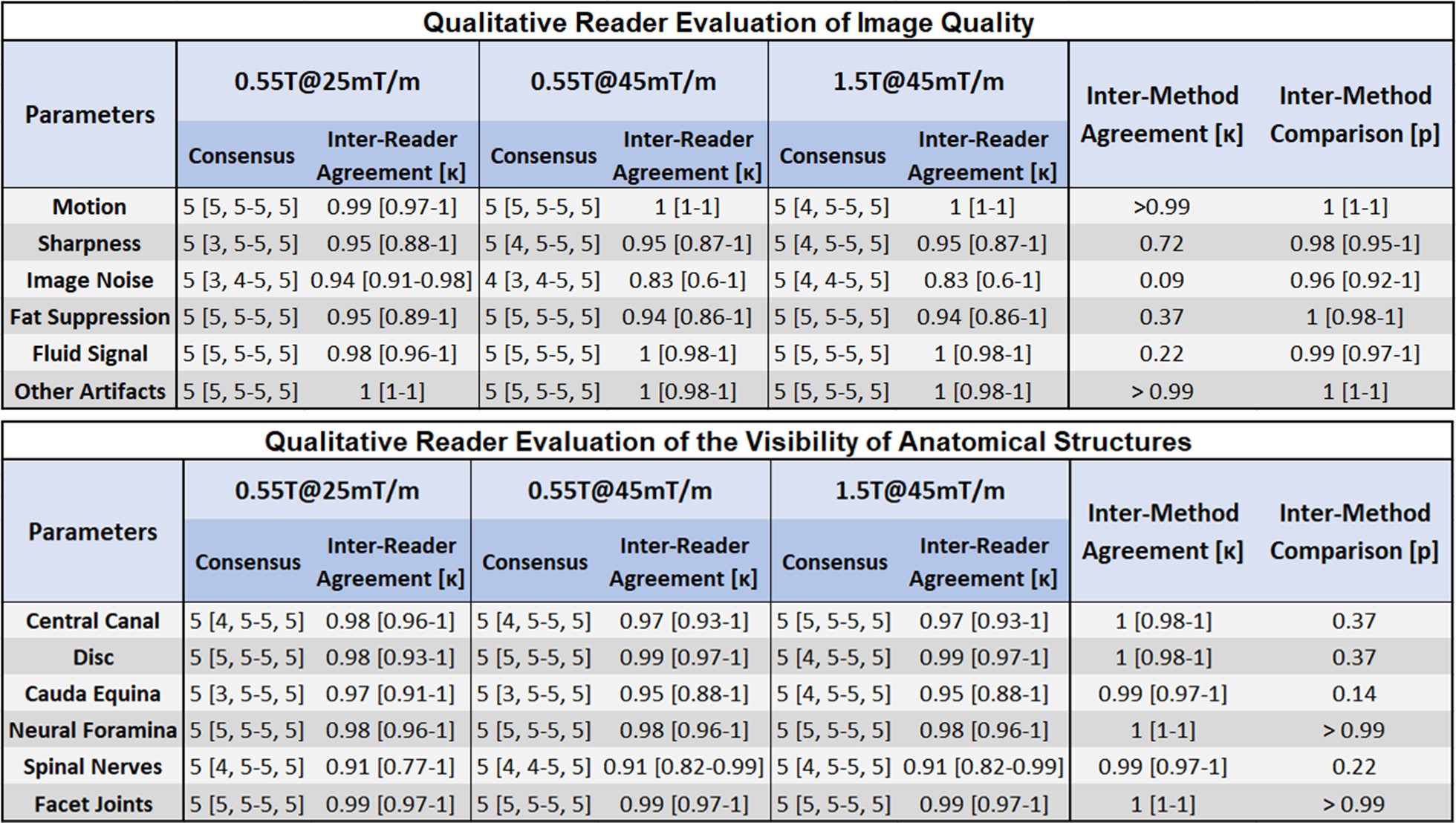
Following IRB approval for prospective data collection and informed consent, ten volunteering participants (7 men, three women; mean age 34.1 ± 9.1 years) were enrolled between March and October 2020. All participants underwent three separate MRI examinations, using a commercial MRI system (1.5T MAGNETOM Aera; Siemens Healthcare GmbH, Erlangen, Germany) modified to operate as a prototype system at 0.55T field strength with two gradient performance levels (25mT/m, 40 T/m/s and 45mT/m, 200 T/m/s), as well as a clinical 1.5T MRI system (MAGNETOM Sola, Siemens Healthcare) with single gradient system (45mT/m, 200 T/m/s).The MRI protocols consisted of sagittal short-tau inversion recovery (STIR), sagittal T1, sagittal T2, and axial T2-weighted turbo spin echo pulse sequences (Table 1). Following anonymization, application of neutral labels, and randomization, three full-time fellowship-trained musculoskeletal radiologists independently evaluated the MRI studies. Quantitative analysis included signal-to-noise (SNR) ratios and contrast-to-noise (CNR) measurements6. Qualitative evaluations included technical image quality assessments (motion, noise, artifacts, edge sharpness of structures, fluid brightness, and fat suppression) using a standardized, five-point Likert scale, as well as visibility of anatomical structures (spinal canal, disc, cauda equina, neural foramen, spinal nerves, and facet joints) using the same Likert scale. All three readers finally ranked corresponding MRI studies in a blinded side-to-side comparison. Statistical evaluations included interrater agreement assessments using kappa statistics and comparison for significant differences using nonparametric tests. P-values of less than 0.5 were considered statistically significant.
Results
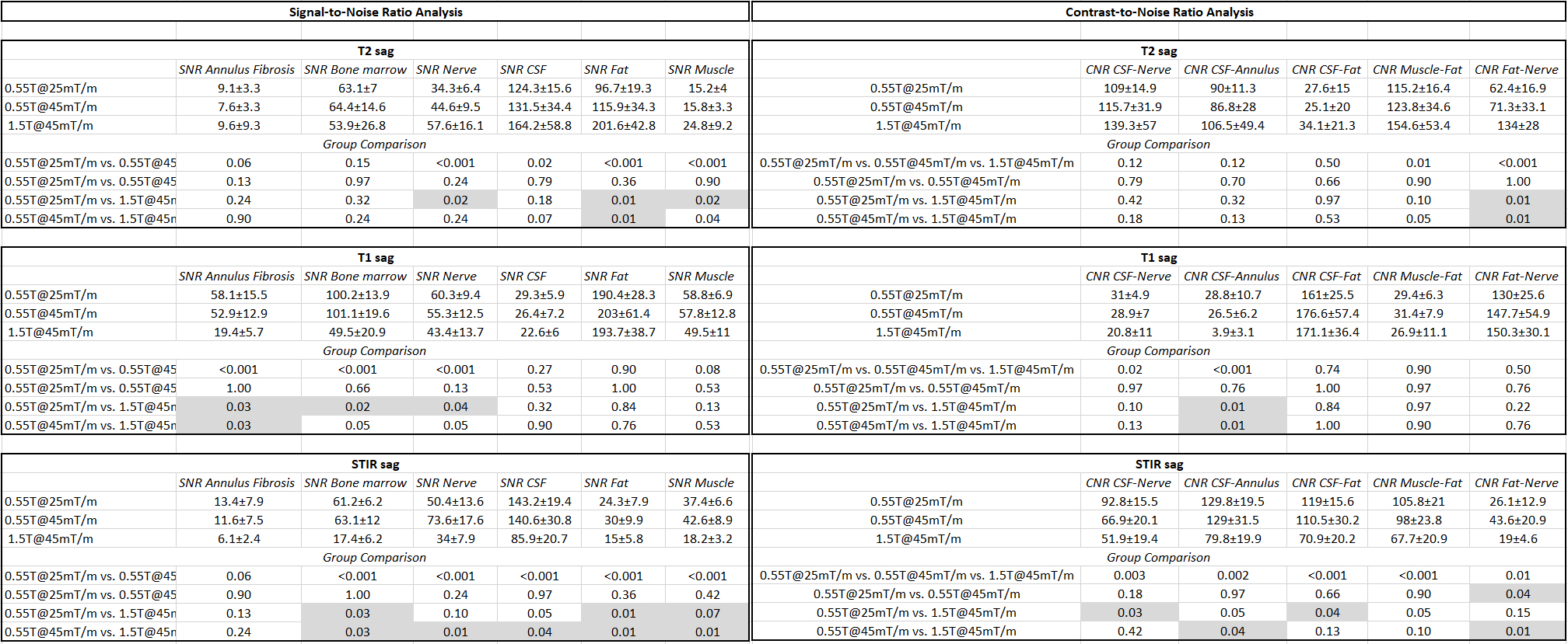
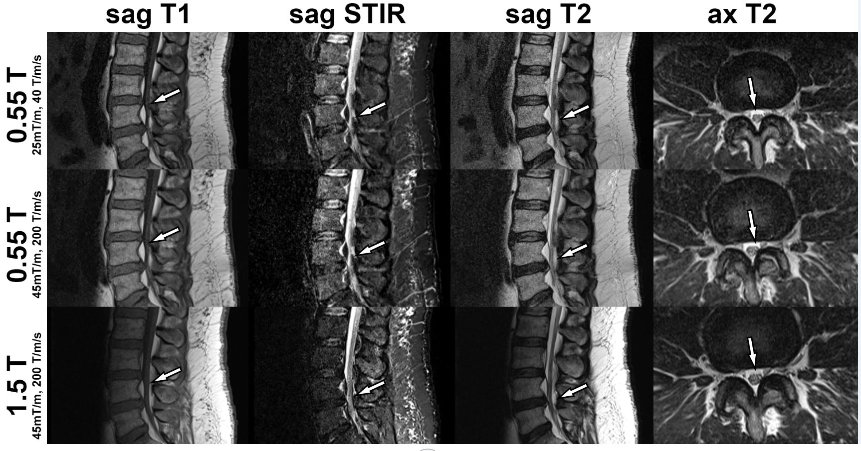
A total of 30 MRI studies were successfully acquired. Regardless of field strength and gradient performance, all MRI studies had sufficient SNR (Table 2) and CNR (Table 2) of critical structures of the spine with small, but in part, statistically significant differences. There was no difference between the two different 0.55T gradient systems. 1.5T MR images showed higher SNR of neural elements on T2-weighted MR images (p<0.001), and lower SNR for bone marrow (p<0.001) and neural elements (p<0.001) on T1-weighted MR images. 1.5T MR images had higher T2 SNR of fluid and lower T1 SNR of skeletal muscle than of 0.55T, although the difference was not statistically significant. 0.55T STIR MR images had higher SNR of bone marrow, neural elements, CSF, fat, and muscle than 1.5-T MRI (p<0.001). Overall CNR of T1- and T2-weighted MR images were similar among all three MRI techniques, whereas 0.55T STIR MR images had wider CNRs at 0.55T than 1.5T. There was good to very good image quality for all three MRI techniques with very good reader agreements (κ≥0.83) and no significant differences (p≥0.09) (Table 3). Similarly, all three MRI techniques showed adequate to very good visibility of anatomical structure with very good readers agreements (κ≥0.91) and no significant differences (p≥0.14) (Table 3). The side-to-side comparison demonstrated no unanimous reader preference between three MRI techniques (p=0.64), indicating all three MRI techniques provided similar image quality (Figures 1 and 2).Discussion
Our study shows that high-performance, low-field-strength 0.55-T MRI of the lumbar spine can produce similarly high image quality and good-to-very good visualization of anatomical structures compared to 1.5T MRI. Advanced low-field MRI systems provide an opportunity to increase accessibility, allowing for longer but clinically feasible acquisition times of approximately 24 minutes, compared to approximately 6 minutes at 1.5. Affordable and accessible MRI has become even more relevant in the light of the most recent economic pressure on health care systems worldwide during and likely after the COVID19 pandemic. Our initial results support the great potential of next-generation low-field MRI systems with substantially lower cost and similar image quality than state-of-the-art clinical 1.5-T MRI systems to detect lumbar spine abnormalities. Further studies are required to validate our results and evaluate the accuracy of 0.55-T MRI in symptomatic patients.Conclusion
High-performance, low-field-strength 0.55-T MRI of the lumbar spine can produce similarly high image quality and good-to-very good visualization of anatomical structures compared to 1.5T MRI.Acknowledgements
The authors would like to acknowledge the assistance of Siemens Healthcare in the modification of the MRI system for operation at 0.55T under an existing research agreement between NYU and Siemens Healthcare.References
1. Runge VM, Heverhagen JT. Advocating the Development of Next-Generation High-Relaxivity Gadolinium Chelates for Clinical Magnetic Resonance. Invest Radiol. 2018;53(7):381-389. doi:10.1097/RLI.0000000000000454
2. Campbell-Washburn AE, Ramasawmy R, Restivo MC, et al. Opportunities in Interventional and Diagnostic Imaging by Using High-Performance Low-Field-Strength MRI. Radiology. 2019;293(2):384-393. doi:10.1148/radiol.2019190452
3. Bendix T, Sorensen JS, Henriksson GAC, Bolstad JE, Narvestad EK, Jensen TS. Lumbar modic changes-a comparison between findings at low- and high-field magnetic resonance imaging. Spine. 2012;37(20):1756-1762. doi:10.1097/BRS.0b013e318257ffce
4. Lee RKL, Griffith JF, Lau YYO, et al. Diagnostic capability of low- versus high-field magnetic resonance imaging for lumbar degenerative disease. Spine. 2015;40(6):382-391. doi:10.1097/BRS.0000000000000774
5. Solgaard Sorensen J, Kjaer P, Jensen ST, Andersen P. Low-field magnetic resonance imaging of the lumbar spine: reliability of qualitative evaluation of disc and muscle parameters. Acta Radiol Stockh Swed 1987. 2006;47(9):947-953. doi:10.1080/02841850600965062
6. Aja-Fernández S, Vegas-Sánchez-Ferrero G, Tristán-Vega A. Noise estimation in parallel MRI: GRAPPA and SENSE. Magn Reson Imaging. 2014;32(3):281-290. doi:10.1016/j.mri.2013.12.001
Figures