3817
Towards Automated Scanning: A Framework for Maintaining Constant SNR and T1-contrast for SPGR and MP-RAGE1Department of Radiology, Mayo Clinic, Rochester, MN, United States
Synopsis
We present a framework for maintaining constant SNR and T1 contrast in SPGR and MP-RAGE scans that employ non-identical scan parameters. Central to this work are the concept of time constant TA and simplified SNR expression. By adjusting the flip angle to keep TA constant and adjusting the number of spiral arms to match SNR, we demonstrated constant SNR and T1 contrast under varied ADC time, TR, and FOV. This work ultimately aims for reducing image variability, automating scanning, reducing technologist intervention, and increasing patient safety.
Introduction
The signal-to-noise ratio (SNR) and tissue contrast are defining characteristics of MR images. In order to standardize image quality and content, automatically maintaining constant SNR and contrast (without technologist intervention) across multiple scans employing non-identical scan parameters remains challenging. For example, changes in the ADC time and repetition time (TR) alter the sampling efficiency and longitudinal relaxation time. These changes, together with other parameters such as scan time and flip angle, affect image SNR and T1 contrast. While such changes are necessary in certain applications, consistent image SNR and contrast may be desired characteristics that benefit precise quantitative studies and clinical diagnosis.Here, we demonstrate a general framework in which image SNR and T1 contrast remain unchanged under different imaging conditions. Using spoiled gradient-echo (SPGR) and MP-RAGE scans that employ spiral trajectories, we demonstrate its efficacy with a particular focus on the changes in the length of the spiral readout trajectory and the corresponding changes in TR. Central to this framework are the concepts of time constant TA and the SNR expression derived for SPGR signals, as explained below.Methods
The time constant TA defines the rate of longitudinal magnetization loss by RF pulses applied each TR in an SPGR experiment, and is related to the flip angle by$$\cos\alpha = e^{-TR/TA}.$$
A previous related work1 demonstrated that T1 contrast stays equivalent among scans where the flip angle and TR are adjusted to keep the TA constant. Further, assuming $$$TR \ll T1$$$ and $$$TR \ll TA$$$ in the signal equation for SPGR, one can show the image SNR in SPGR scans is proportional to the total scan time T and ADC time $$$\tau$$$:
$$SNR\sim\sqrt{T\tau}S_{\text{avg}}$$
where Savg is the sampling density correction weights. Thus, for scans employing different lengths of readout trajectories (measured by the ADC time $$$\tau$$$), the number of spiral interleaves can be adjusted to control the scan time T and match SNR per Eq. 2.
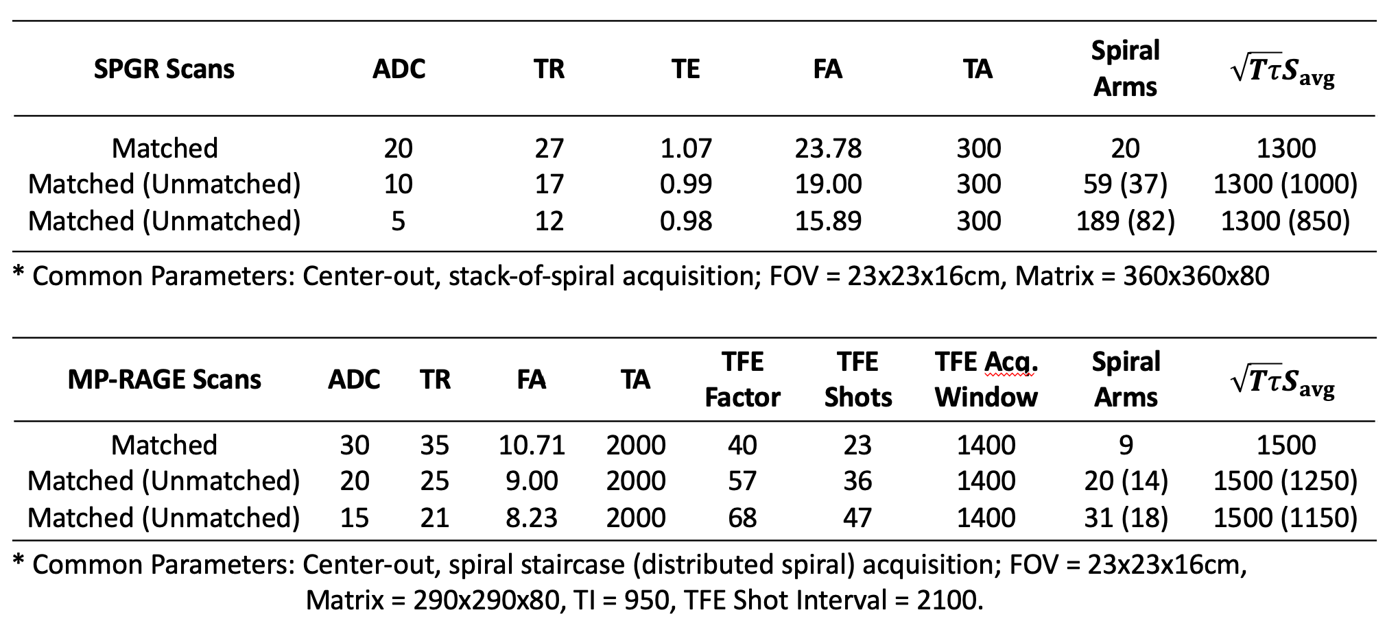
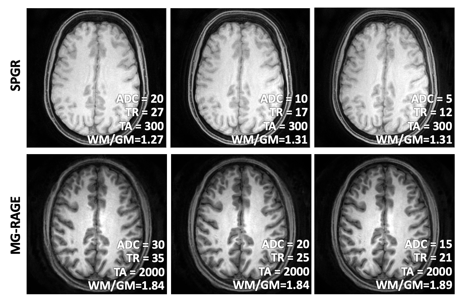
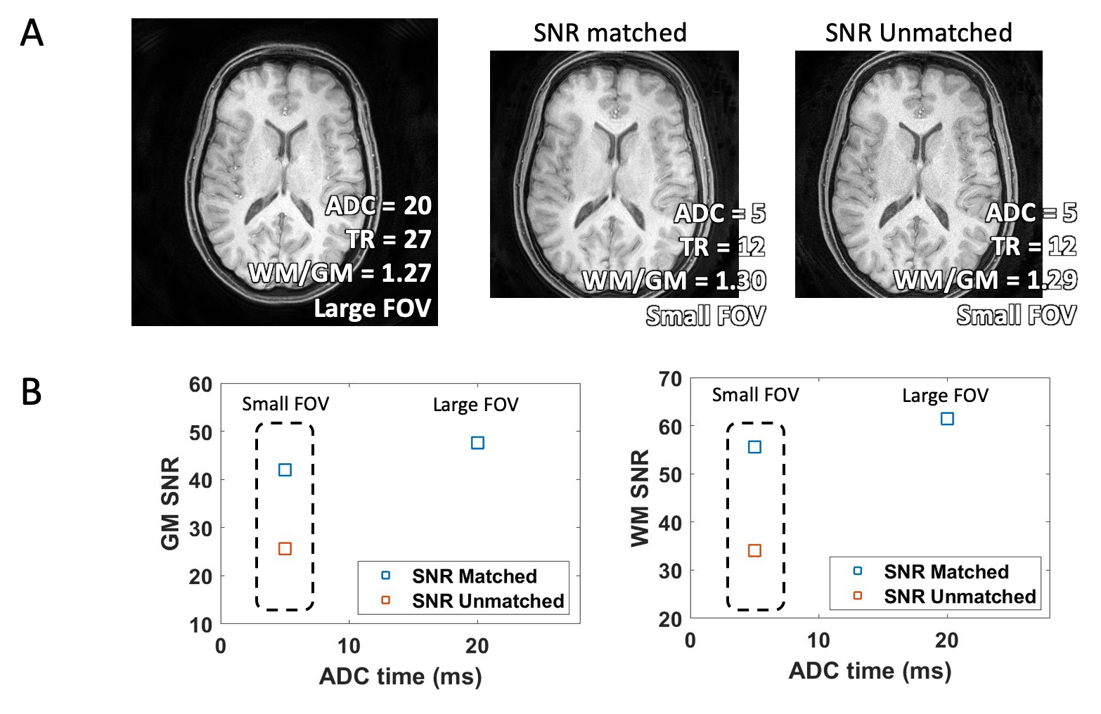
Using a 3T MRI scanner (Philips Ingenia Elition X) and a 14-channel head coil, 3D stack-of-spiral SPGR scans and 3D spiral-staircase2 MP-RAGE3 scans were acquired in 2 volunteers and 1 volunteer, respectively. Each scan was acquired using three different lengths of spiral readout trajectories with varied ADC time ($$$\tau$$$) and TR. The change in TR was accompanied with flip angle adjustment to keep TA constant and maintain equivalent T1 contrast. Also, the number of spiral arms was adjusted to keep $$$\sqrt{T\tau}S_{\text{avg}}$$$ constant and match SNR among the scans having different spiral trajectory lengths. For comparison, reference scans were obtained with the minimum number of spiral interleaves required to support the FOV. In one volunteer, SPGR scans were acquired with two different FOVs, while the number of spiral arms in the small-FOV scan was adjusted to match the SNR of the large-FOV scan. The same small-FOV scan was acquired without spiral arm adjustment as a reference for the SNR-unmatched case. The flip angle was also adjusted to keep TA constant, as the small and large FOV scans employed different ADC times. Detailed scan parameters are listed in Table 1.The SNR was measured at an identical axial slice separately in gray and white matter, which were automatically segmented using the FMRIB Software Library (FSL)4. The noise was measured by 2X-oversampling each readout, and then subtracting the images reconstructed from even- and odd samples.
Results
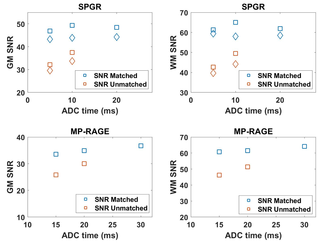
By keeping the TA constant in scans having different ADC times, both SPGR and MP-RAGE images yielded equivalent T1 contrast despite having different TR’s (Figure 1). The constant T1 contrast was measured in terms of WM/GM signal ratio, included in Figure 1. With the number of spiral arms adjusted to match the SNR expression (Eq. 2), both gray and white matter SNR stayed equivalent across the three ADC times examined in each scan type ($$$\tau$$$=5,10,15ms in SPGR, $$$\tau$$$=15,20,30ms in MP-RAGE), as shown in Figure 2. When these scans employed unadjusted number of arms with minimal Nyquist sampling to support the FOV (SNR-unmatched), these reference scans exhibited substantial SNR decrease for shorter trajectory lengths, as expected. The two scans having different FOVs agreed in SNR within 10%, when the number of spiral arms was adjusted to match SNR. In comparison, the SNR-unmatched, smaller-FOV scan dropped 45% in SNR without such adjustment.Discussion
In this work, we utilized the concept of TA and SNR to demonstrate the framework of realizing constant T1 contrast and image SNR when spiral trajectory lengths, TR, and FOV varied in SPGR and MP-RAGE scans. While demonstrated with spiral scans, this framework has general applicability that the same concepts can be applied towards other readout trajectory types such as Cartesian, radial, etc. However, certain trajectory types2,5 are naturally well suited for adjusting the number of readout shots to match the SNR (i.e. distributed spiral sampling). Given the efficiency and flexibility of trajectory patterns, we felt that it was most adequate to demonstrate this framework using spiral scans. The ultimate goal of this work is to more fully automate the scanning procedure so that technologists can interact more with the patient and less with the scanner, increasing patient safety and improving patient experience, while simultaneously reducing image variability.Acknowledgements
This work was partially funded by Philips Healthcare.References
1. Pipe JG and Robison RK. Simplified Signal Equation for Spoiled Gradient Echo MRI. Proceedings of the 19th Annual Meeting of ISMRM, Stockholm, Sweden.
2. Anderson AG 3rd, Wang D, Pipe JG, Controlled aliasing for improved parallel imaging with a 3D spiral staircase trajectory. Magn Reson Med. 2020 Aug; 84 (2):866-872 Epub 2020 Jan 22
3. Brant-Zawadski M, Gillan GD, Nitz WR. MP RAGE: a three-dimensional, T1-weighted, gradient-echo sequence—initial experience in the brain.
4. FMRIB Software Library (FSL), FMRIB Centre, University of Oxford, UK.
5. Turley DC and Pipe JG. Distributed spirals: A new class of three-dimensional k-space trajectories. Mag Res Med. 2012; 70(2):413-419
Figures