3809
Accelerated hyperpolarized 13C MRI using 23Na coil sensitivity mapping, proof of concept1Physiology, Anatomy, and Genetics, University of Oxford, Oxford, United Kingdom, 2Oxford Centre for Magnetic Resonance, University of Oxford, Oxford, United Kingdom, 3Radiology, Oxford NHS Foundation Trust, Oxford, United Kingdom, 4Health Technology, Danish Technical University, Copenhagen, Denmark, 5Aarhus University, Aarhus, Denmark, 6Danish Technical University, Copenhagen, Denmark
Synopsis
Multi-channel acceleration of hyperpolarised 13C MRI is hindered by a lack of prior coil sensitivity profile knowledge. Here we harness a 13C tuned flexible multi-channel array to provide subject specific coil sensitivity measurements from 23Na and subsequent SENSE reconstruction of under-sampled hyperpolarised imaging data.
Introduction
Hyperpolarized 13C MRI is a developing clinical technique, with potential applications in numerous pathologies (1–3). However, the clinical use of flexible multi-channel receive coils is hampered by the unknown sensitivity profile prior to acquisition. This, in turn, inhibits both the spatial sensitivity correction of acquired data, as well as partial phase field of view acceleration for imaging.It has already been shown that the sodium-23 (23Na) resonance can provide a useful prescan for system calibration prior to imaging due to their similar gyromagnetic ratios and high natural abundance in vivo (4). Here we extend this work using a novel flexible multi-channel receive coil tuned to 13C resonance frequency, but with similar decoupling characteristics at both 13C and 23Na frequencies. This ensures similar spatial sensitivity profiles, enabling convenient workflow for undersampling strategies such as SENSE reconstruction of thermal and hyperpolarized 13C imaging data by relying on the 23Na spatial profiles.Methods
All scanning was performed at 3T (GE MR750, GE Healthcare, WI).Phantom studies
The 8-channel receive 13C coil was wrapped around a saline/pyruvate phantom (4) with a clamshell exciter system (Rapid, Rimpar).23Na 2D Chemical Shift Imaging (CSI) (Flip angle (FA) = 45 degrees, repetition time (TR) = 250 ms, echo time (TE) = 4 ms, Field of view (FOV) = 280mm2, slice thickness = 30mm, in-plane resolution = 10mm2, bandwidth = 5kHz, points = 1024, Averages (NEX) = 1) and 13C 2D CSI were performed (FA = 90 degrees, TR = 2500 ms, TE = 4 ms, Field of view (FOV) = 280mm2, in-plane resolution = 10mm2, bandwidth = 5kHz, points = 1024, NEX = 4).
2D spiral imaging (2 arms, TE = 2/4 ms, NEX (13C/23Na) = 64/150, imaging time (13C/23Na) = 3 hours/90 seconds, reconstruction matrix = 128x128, all other parameters matched to 2D CSI) was also used to map channel sensitivity at both 13C and 23Na frequencies. Fully sampled and reduced phase field of view 2D 23Na cartesian data were acquired (FOV/acquisition matrix/reconstruction matrix =280x280/28x28/56x56, 280x140/28x14/56x28, TE = 4 ms, TR = 40 ms, flip angle = 40 degrees, NEX = 16).
In vivo study
A Danish domestic pig was anesthetised using Propofol and Fentanyl and mechanically ventilated with 30% oxygen for a target end-tidal CO2 of 5.5 kPa.After an injection of hyperpolarised [1-13C]pyruvate, imaging data were acquired using a selective excitation echo planar imaging (EPI) sequence using a reduced phase field of view (FOV = 280 mm, slice thickness = 30 mm, acquisition matrix = 16x8, TE = 23 ms, TR per metabolite = 1 s, total time resolution = 4s, flip angles (Pyr/Lac/Bicarb = 10,40,40, respectively, total time points = 15). 2D channel sensitivity imaging was performed using a 2D 23Na spiral acquisition as above.
Image reconstruction and processing
All images were processed in Matlab (The Mathworks, 2019b). 2D CSI images were reconstructed using singular value decomposition coil combination and the peak signal intensity of the phased real signal component in each voxel used to form images. 2D spiral images were reconstructed using gridding and a fast Fourier transform.Under sampled 23Na 2D cartesian and 13C echo planar data were combined using both root sum of squares (RSS) and SENSE(5). SENSE was performed using the complex post-gridding 2D spiral imaging data as coil sensitivity maps.
Results
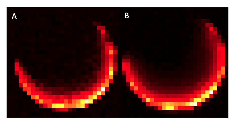
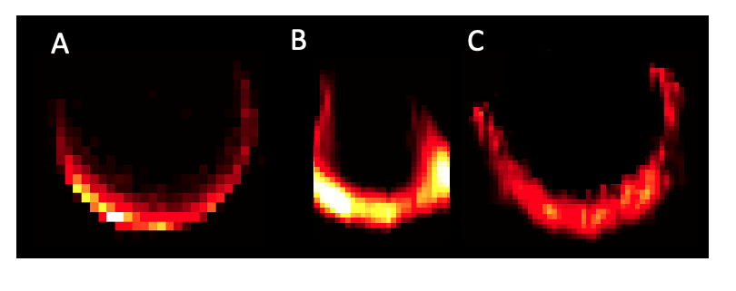
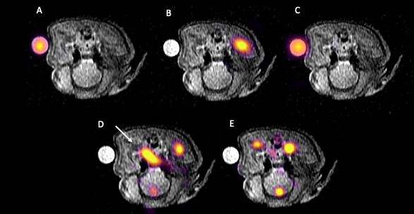
2D CSI showed similar spatial penetration and signal distribution after SVD combination at both 23Na and 13C frequencies (see Figure 1A and B, respectively).Reconstruction of fully sampled and 50% phase field of view cartesian data with RSS and SENSE 23Na data are shown in Figure 2 A, B, and C, respectively. Reconstructions of summed EPI data are shown in Figure 3 (A, B, C, = fully sampled, RSS, and SENSE reconstructions of thermal 13C bicarbonate, respectively. D, E = RSS and SENSE reconstructions of summed [1-13C] pyruvate, respectively), showing good registration of imaging data with both phantom and the vascular distribution of 13C pyruvate after SENSE.
Discussion
These initial results demonstrate the potential for a novel 13C multi-channel flexible array, with similar sensitivity profiles at 23Na, to be used for hyperpolarised imaging, with SENSE based reconstructions. Unfolded 23Na and 13C data show strong similarity to fully sampled imaging, and the in vivo hyperpolarised imaging shows a similar [1-13C]pyruvate bio-distribution in the porcine vasculature to previously published work(6).Channel spatial sensitivity mapping was limited to 2D spirals, but could be extended to faster volumetric 3D ultrashort echo time trajectories such as 3D radial or cones(7). In turn, sensitivity maps could be combined with real-time undersampling such as compressed sensing strategies, to assist in large field of view acquisitions.Further validation and development of flexible 13C/23Na coils will include improved penetration and use in clinical studies on systems provided by all of the main vendors involved in 13C acquisitions.
Conclusion
We have developed a novel flexible multi-channel receive coil that shares similar spatial sensitivity profiles for both the 23Na and 13C resonances and here we show its utility for unwrapping folded hyperpolarized 13C data using SENSE.Acknowledgements
James Grist acknowledges support from the BHF Centre of Research Excellence, University of Oxford.References
1. Chaumeil MM, Larson PEZ, Woods SM, et al. Hyperpolarized [1-13C] glutamate: A metabolic imaging biomarker of IDH1 mutational status in glioma. Cancer Res. 2014;74(16):4247–4257. doi: 10.1158/0008-5472.CAN-14-0680.
2. Hansen ESS, Stewart NJ, Wild JM, Stødkilde-Jørgensen H, Laustsen C. Hyperpolarized 13C,15N2-Urea MRI for assessment of the urea gradient in the porcine kidney. Magn Reson Med. 2016;76(6):1895–1899. doi: 10.1002/mrm.26483.
3. Grist JT, Miller JJ, Zaccagna F, et al. Hyperpolarized 13C MRI: A novel approach for probing cerebral metabolism in health and neurological disease. J Cereb Blood Flow Metab. 2020; doi: 10.1177/0271678X20909045.
4. Grist JT, Sánchez-heredia ESSHJD, Mclean MA, et al. Creating a clinical platform for carbon-13 studies using the sodium-23 and proton resonances. Magn Reson Med. 2020;00(1):1–11. doi: 10.1002/mrm.28238.
5.Pruessmann KP, Weiger M, Scheidegger MB, Boesiger P. SENSE: sensitivity encoding for fast MRI. Magn Reson Med. John Wiley & Sons, Inc.; 1999;42(5):952–962. doi: 10.1002/(SICI)1522-2594(199911)42:5<952::AID-MRM16>3.0.CO;2-S.
6. Miller JJ, Grist JT, Serres S, et al. 13C Pyruvate Transport Across the Blood-Brain Barrier in Preclinical Hyperpolarised MRI. Sci Rep. 2018;8(1):15082. doi: 10.1038/s41598-018-33363-5.
7.Grist JT, Riemer F, McLean MA, et al. Imaging intralesional heterogeneity of sodium concentration in multiple sclerosis: Initial evidence from23Na-MRI. J Neurol Sci. 2018;387:111–114. doi: 10.1016/j.jns.2018.01.027.
Figures