3787
Phase Dispersion from Steady-State Signal Behavior in Phase-Sensitive Multiband Imaging with Application to MREIT1Department of Biochemistry and Molecular Biology, University of Florida, Gainesville, FL, United States, 2School of Biological and Health System Engineering, Arizona State University, Tempe, AZ, United States, 3University of Minnesota, Minneapolis, MN, United States, 4MR R&D Collaboration, Siemens Medical Solutions USA, Atlanta, GA, United States, 5Department of Radiology, Johns Hopkins University, Baltimore, MD, United States
Synopsis
Incorporating multiband excitation into phase-sensitive imaging introduces distortions in the phase measurements. We used simulations and phantom images to show that a previously unreported phase dispersion problem arises within multiband-slice phase maps. We show that this problem can be understood in terms of the steady-state signal behavior and propose imaging protocols to resolve it. Human scans using these protocols show minimal phase dispersion. We are adopting this protocol for our studies of phase-sensitive magnetic resonance electric impedance tomography (MREIT).
Introduction
Multiband excitation for simultaneous multi-slice (SMS) imaging1 can be incorporated into MREIT measurements2 to allow whole brain coverage, and speed up current density mapping during transcranial electrical current stimulation. In multiband excitation, the phase of the multiband radiofrequency (RF) pulse is modulated for each slice using a frequency-offset, phase gradient to shift the slice in the phase-encoding direction. For a given multiband factor, MB, the field of view, FOV, in the phase-encoding direction is chosen so that $$$FOV = (MB \times FOV_{object})/R$$$, where $$$FOV_{object}$$$ is the desired object FOV, and aliasing is controlled by the reduction factor, $$$R$$$. The phase of each slice is modulated so that the slice $$$s$$$ is shifted by $$$\triangle y_{s}=(s-1)\times(FOV_{object}/R)$$$ in the phase-encode direction. This is achieved by adding the multiband phase $$$\varnothing_{MB}(m)=m\triangle k_{y}\triangle y_{s}$$$ to the RF pulse for each slice at each $$$\triangle k_{y}$$$phase-encode step, $$$m$$$.1 For MREIT, phase measurements are performed with a relatively short TR, so the added RF pulse phase,$$$ \varnothing_{MB}$$$, can interfere with RF spoiling and disturb steady-state formation. In the present study, simulations of steady-state performance are presented and compared to measurements in phantom and human brain images. We show that a previously unreported phase dispersion problem within multiband slices arises in phase measurements, which can be understood based on the signal's behavior when approaching steady state. We then show that this problem can be resolved using different imaging protocols.Methods
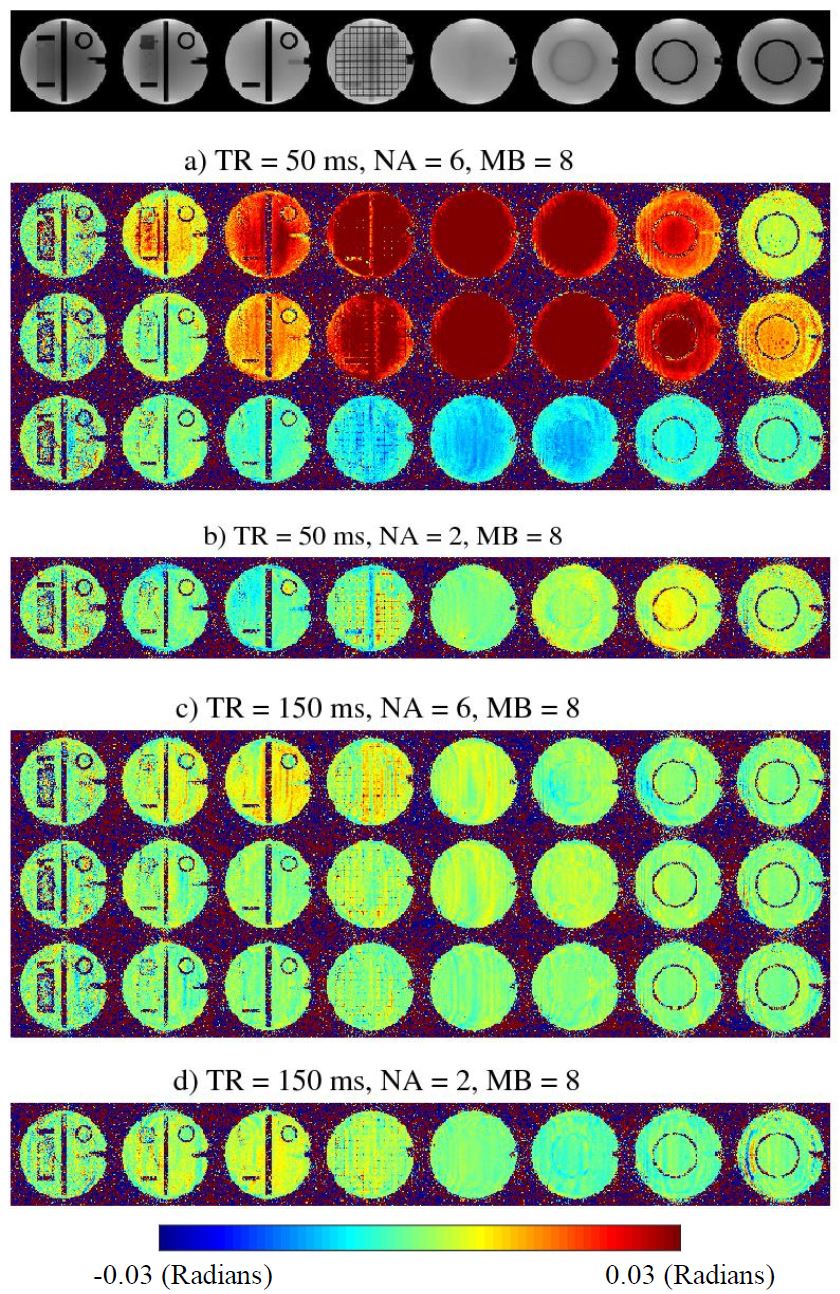
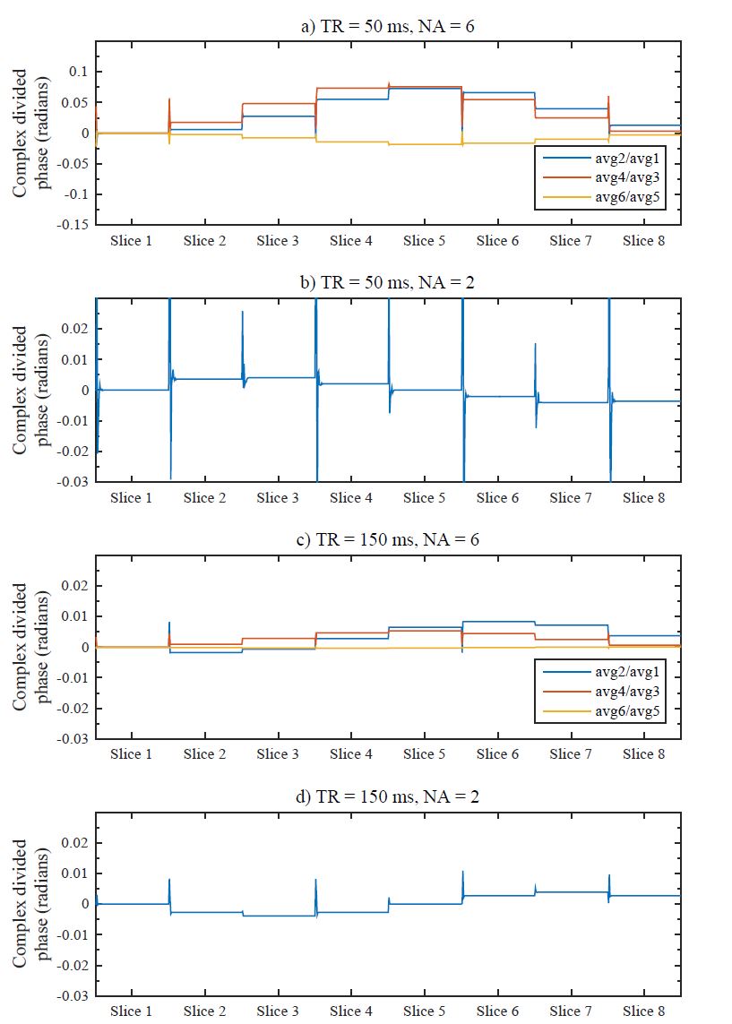
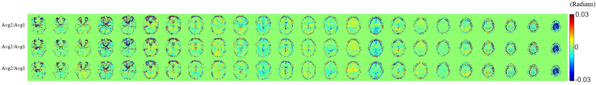
Simulations: Signal magnitude and phase approach to steady-state was simulated at each repetition step, $$$n$$$, before and after applying each RF pulse3 and the multiband phase at each phase encode step, $$$m$$$, was added to the phase of the RF pulse. Relaxation-time dependent transverse and longitudinal states were calculated at the end of each TR interval. The phase of the RF pulse at each step is given by $$$ \varnothing_{RF}(n,m)=\varnothing_{Spoiler}(n)+\varnothing_{MB}(m)$$$ , where $$$\varnothing_{Spoiler}(n)=\frac{1}{2}n(n-1)\psi_{0}$$$ with $$$\psi_{0}$$$ being the RF spoiling phase increment. This phase was added after the signal was updated at each RF pulse in the simulation. The phase of the measured signal at each step was calculated after applying the RF pulse.Imaging Experiments: An American College of Radiology (ACR) phantom was scanned on a 3T system (MAGNETOM Prisma, Siemens Healthcare, Erlangen, Germany) using a prototype multi-band gradient-echo (GRE) sequence. Multiband excitation was incorporated into the multi-echo, GRE sequence which we use for MREIT. We used two and six numbers of averages (NA) and TR=50 ms (single multiband slice excitation, repeated 3 times) and TR=150 ms (3 interleaved multiband slice excitation) for scans with MB=8. The following parameters were used for all scans: FOV=$$$(MB \times FOV_{object})$$$, FOVobject=224x224 mm2, matrix size=100x100, slice thickness=5mm, Nslices=24, Necho=10, TE1=7 ms, ∆TE=3 ms, flip angle=30. Simulations were performed using the same imaging parameters as the imaging experiments and relaxation times of the ACR phantom. A human volunteer was scanned on the Phillips Ingenia 3T scanner using the same parameters except for FOV=$$$(MB \times FOV_{object})/2$$$.4 Phase maps were calculated for the phantom and human scans, without any current injection by complex dividing consecutive pairs of averages in each scan2.
Results
Results from ACR phantom measurements on the and corresponding simulations are shown in Figs. 1 and 2 respectively. It can be seen that a large phase dispersion dominates phase maps when using TR=50ms and NA=6 (see Fig. 1, a), which is significantly reduced using TR=150 ms or NA=2 (see Fig. 1, c). The simulations in Figure 2 show the phase of the signal after complex-dividing consecutive averages and show excellent agreement with phantom images. Fig. 3 shows the results of human brain scans using TR=150ms and NA=2.Discussion
Using a longer TR results in less residual magnetization and improves the phase maps across the imaging volume. The effect of number of averages can be understood based on the steady-state signal's behavior and the order of averaging in MREIT scans. In MREIT measurements, all averages are acquired for each phase-encode step in order to allow current reversal for each average as close in time as possible to eliminate any phase changes due to sources other than injected current. Because of the phase added to the RF pulse at each phase-encode step in multiband measurements, this order of acquisition results in violation of one of the conditions for producing steady states, i.e., that the phase of the RF pulse must change according to a second-degree polynomial.5,6 The phase of the RF pulse for multiband measurements can be rewritten as $$$\varnothing_{RF}(n)=\frac{1}{2}n(n-1)\psi_{0}+\lfloor\frac{n}{NA}\rfloor\triangle k_{y}\triangle y_{s}$$$, where $$$\lfloor.\rfloor$$$ denotes the floor function, which shows the deviation from second-degree polynomial behavior for multi-average acquisition of each individual slice and how this deviation is affected by the number of averages.Conclusion
Incorporating multiband acquisition into phase-sensitive image measurements results in phase dispersion within multiband slices due to the way in which phase manipulation of multiband RF pulse interferes with the performance of RF spoiling and formation of a steady-state. Using a longer TR value (here 150 ms instead of 50 ms) and combining three two-average scans, instead of acquiring one six-average scan, almost completely removed the phase dispersion from the resulting phase maps. This protocol will be adopted in our future studies of MREIT.Acknowledgements
This work was supported by NIH award, RF1MH114290, and in part by an NIH award, S10 OD021726, for High End Instrumentation. A portion of this work was performed in the McKnight Brain Institute at the National High Magnetic Field Laboratory’s Advanced Magnetic Resonance Imaging and Spectroscopy (AMRIS) Facility, which is supported by National Science Foundation Cooperative Agreement No. DMR-1644779 and the State of Florida.References
1. Breuer, F. et al. Controlled aliasing in parallel imaging results in higher acceleration (CAIPIRINHA) for multi-slice imaging. Magnetic Resonance in Medicine. 2005;53(3):684-691.
2. Kasinadhuni, A. K. et al. Imaging of Current Flow in the Human Head During Transcranial Electrical Therapy. Brain Stimulation. 2017;10(4):764-772.
3. Scheffler, K. A Pictorial Description of Steady-States in Rapid Magnetic Resonance Imaging. Concepts in Magnetic Resonance. 1999;11(5):291-304.
4. Chauhan, M. et al. Current Density Measurements in the Human Brain in-vivo during TES treatment, using Multi-Bandmethods Proc. Intl. Soc. Mag. Reson. Med. 28 (2020),3179.
5. Zur, Y. et al. Spoiling of Transverse Magnetization in Steady-State Sequences. Magnetic Resonance in Medicine. 1991;21(2):251-263.
6. Sobol, W. T. and Gauntt, D. M. On the Stationary States in Gradient Echo Imaging. Journal of Magnetic Resonance Imaging. 1996;6(2):384-398
Figures