3762
aDWI-BIDS: Advanced Diffusion Weighted Imaging Metadata for the Brain Imaging Data Structure1School of Physics and Astronomy, Cardiff University, Cardiff, United Kingdom, 2CUBRIC, Cardiff University, Cardiff, United Kingdom, 3Universidad de Valladolid, Valladolid, Spain, 4Department of Diagnostic Radiology, Lund University, Lund, Sweden, 5Image Sciences Institute, University Medical Center Utrecht, Utrecht, Netherlands, 6Harvard Medical School, Boston, MA, United States
Synopsis
We present an extension to the Brain Imaging Data Structure (BIDS) to specialise it for diffusion weighted imaging. Detailed attribution of experimental parameters to regions of an aquisition is made possible with plain text files which remain compliant with BIDS. Complex diffusion encoding, slice-level diffusion encoding, and data collected with varying experimental parameters throughout the acquisition are all supported. Scope exists for reporting on RF pulses and gradient pulses in general within the sequence, without restriction to diffusion pulses.
Body of Abstract
Quantitative MRI studies require an exhaustive modality-dependent list of parameters along-side the imaging data. The Brain Imaging Data Structure (BIDS) is rapidly gaining traction in the brain imaging community for organising data in a coherent and standardised fashion1. However, not all modalities are specified exhaustively within BIDS. Developments in diffusion encoding methods expose limitations in how the BIDS provides metadata about the diffusion encoding used throughout a dataset. Datasets employing complex diffusion encoding2–8 may vary numerous sequence parameters throughout an acquisition, necessitating tabular recording of this variation throughout the dataset2. A generic, BIDS compliant scheme to record the encoding employed within a dataset would facilitate its automated analysis, as well as permitting a standardised format for recording archive-quality data.We considered a wide set of experimental and established diffusion imaging paradigms and assessed which metadata were necessary for subsequent quantification / interpretation of magnetic resonance images. This was carried out by examination of current non-BIDS metadata standards proposed1, 9, 10, as well as component level analysis of the encoding methods used11. This incorporated expectations for pre- and post-processing, heuristics for pipeline automation, sequence visualisation, modality comparison, and ingestion for large mixed-modality datasets for quantitative MRI.
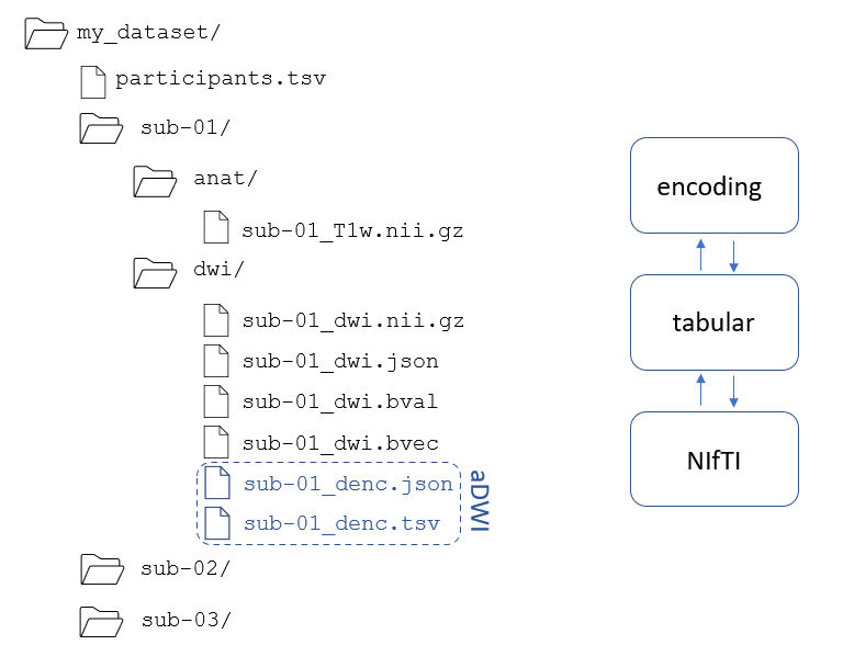
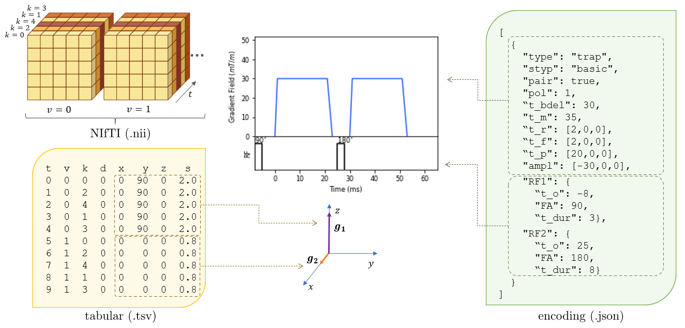
A recording scheme was realised for comprehensive description of diffusion metadata, and is also appropriate for describing other areas variable or experimental gradient or RF activity. This was expressed as an iterable structure, comprised of three files: a NIfTI10 (.nii(gz)) file for multidimensional imaging data, an encoding file (.json) conveying the diffusion encoding used, and a tabular (.tsv) file to detail how the diffusion encoding varies throughout the multidimensional NIfTI (figure 1).
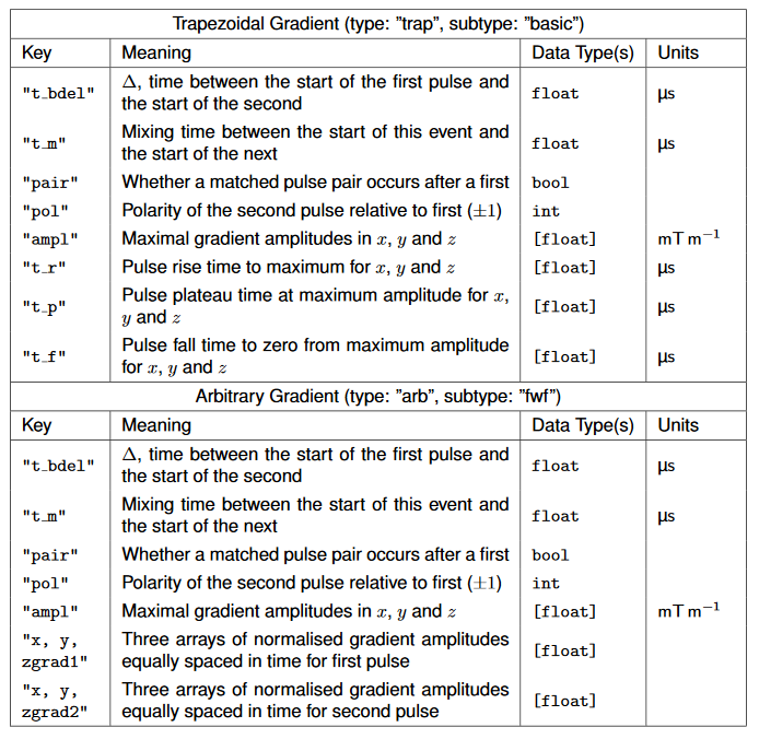
The encoding file uses an object-oriented approach to describe a collection of ordered lists of sequence objects (”events”). Multiple types of encoding can be expressed in one file. Events are chained together to provide a description of a region of a sequence. Events are modular, and can contain terse or detailed descriptors without loss of functionality. Moreover, they may be programmatically specified using JSON schema12 (table 1), and hence automatically recognised and handled. We make a repository of JSON schemas and other tools for gradient events available on our GitHub (https://github.com/JAgho/aDWI-BIDS). Methods are specified to express arbitrary gradient waveforms, as well as most common gradient pulses.
The tabular file divides acquisitions within the NIfTI into rows, where acquisitions may be either a single slice, or a single volume from the NIfTI (figure 2). The columns may refer to any parameters we aim to describe, allowing the tracking of changes to parameters between acquisitions. As well, they may refer to objects within the encoding file. This permits the attribution of objects to every acquisition, where the objects convey the type and parameters of the diffusion encoding employed.
Additionally, we may use the tabular file to communicate variations within an encoding object. This permits variation in a sequence to be given for each slice or volume in a dataset. This simplifies experiments examining the variation of a single sequence parameter, and permits variable encoding used from slice to slice to be recorded.
The structure proposed is flexible, and can describe many experimental conditions. Rich metadata labels components of a scan with their respective parameters. Consequently heterogenous NIfTIs (using multiple diffusion encodings) can be handled differently according to their properties. It is common to include multiple acquisition settings within a single NIfTI, e.g. different b-values. These are clearly identified within the new structure, without reliance on .bval files.
In vivo scans are complicated by interruptions, subject motion, and many other factors. Correction of artifacts arising from complications, and analyses to derive diffusion measures, relies on the ability to group metadata, and to merge and split datasets freely. Combined analyses like diffusional variance decomposition13 may need merged datasets, or recognition of a single b=0 image as registration for a subset of data. Free labelling of regions of a combined NIfTI’s meta-data simplifies pipeline design, incorporating data for combined processing steps, and extraction of data subsets based on metadata properties.
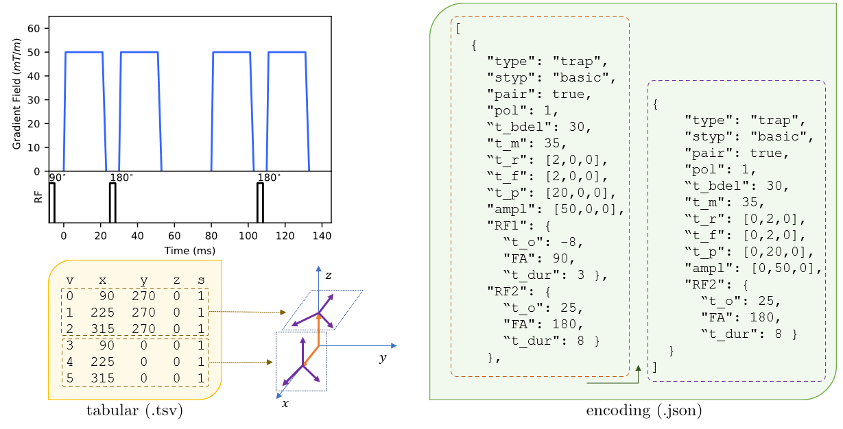
aDWI-BIDS also conveys multiple scan directions without repetition of an underlying diffusion pulse sequence. By providing a set of Euler angles alongside each volume or slice, we permute a prototypical encoding object into a collection of unique rotated forms (figure 3).
Finally, the scheme is easily extended to other sequence facets. Behaviours like shaped or multi-channel RF pulses may be recorded with positions within a sequence given unambiguously. This utility applies to readout behaviour as well, and waveform-based readout gradients may be given in totality. Should distortion or phase correcting approaches be developed to address artefacts in recorded data, this may be applied retroactively, with full knowledge of the pulse sequence that produced it.
The scheme proposed allows the recording of the underlying diffusion experiment in an acquisition in full detail. If new approaches are developed to interpret or process diffusion data, these may be applied to historical data in a modular way, simplifying the development of processing pipelines, and allowing ingestion of data acquired with multiple acquisition parameters. This has utility in, e.g., machine learning applications, large population studies, data harmonisation14 or in multi-modal microstructure methods.
Acknowledgements
This work was supported by the Science and Technology Facilities Council, UK, through grants ST/00209X/1 (MIDaC) and Impact Acceleration Account (MISP, Cardiff University).
We would also like to thank Andreas Papageorgiou of the Cardiff University School of Physics and Astronomy for their assistance with pipeline planning and containerisation of software.
More generally, thanks to the team at CUBRIC for their support, patience, and training.
References
1. Gorgolewski, K. J. et al. Scientific Data 2016, 3, 160044.
2. Szczepankiewicz, F.; Westin, C.-F.; Nilsson, M. Journal of Neuroscience Methods 2020, 109007.
3. Szczepankiewicz, F.; Sjolund, J.; Stahlberg, F.; Latt, J.; Nilsson, M. PLOS ONE 2019, 14, Publisher: Public Library of Science, e0214238.
4. Shemesh, N.; Ozarslan, E.; Komlosh, M. E.; Basser, P. J.; Cohen, Y. NMR in Biomedicine 2010, 23, 757–780.
5. Shemesh, N. et al. Magnetic Resonance in Medicine 2016, 75, 82–87.
6. Eriksson, S.; Lasic, S.; Topgaard, D. Journal of Magnetic Resonance 2013, 226, 13–18.
7. Merboldt, K.-D.; Hanicke, W.; Bruhn, H.; Gyngell, M. L.; Frahm, J. Magnetic Resonance in Medicine 1992, 23, 179–192.
8. Baron, C. A.; Beaulieu, C. Magnetic Resonance in Medicine 2014, 72, 726–736.4
9. Markus Nilsson, Filip Szczepankiewicz Multi-Dimensional Diffusion MRI Toolbox.
10. Jenkinson, M. NIfTI-1 Data Format — Neuroimaging Informatics Technology Initiative.
11. Bernstein, M. A. Handbook of MRI pulse sequences; King, K. F., Zhou, Z. J., Eds.; Amsterdam: Amsterdam, 2004.
12. Wright, A.; Andrews, H.; Hutton, B.; Dennis, G. JSON Schema: A Media Type for Describing JSON Documents; Internet-Draft; Internet Engineering Task Force, 2019, 75 pp.
13. Szczepankiewicz, F.; van Westen, D.; Englund, E.; Westin, C.-F.; Stahlberg, F.; Latt, J.; Sundgren, P. C.; Nilsson, M. NeuroImage 2016,142, 522–532.
14. Tax, C. M. et al. NeuroImage 2019, 195, 285–299
Figures