3748
Automated scan plane planning for multiple examination parts by modular algorithm developing method1Healthcare Business Unit, Hitachi, Ltd, Tokyo, Japan
Synopsis
Generally, automated scan plane planning methods require the recognition of different landmarks depending on the examination parts. Therefore, algorithms must be tailored to each examination part. In this study, we have proposed a modular algorithm developing method for automatic scan plane planning. In this method, an algorithm is composed of several common processes, and by simply changing the combination of those processes, it can be tailored to different examination parts.
Purpose
Automated scan plane planning is expected to improve MRI scanner usability and provide consistent scan plane prescriptions, which are useful for follow-up examinations. We previously proposed an automated scan plane planning method for brain and spine using 2D multi-slice orthogonal three-plane scout images1,2. Generally, automated scan plane planning methods require the recognition of different landmarks depending on the examination parts. Therefore, algorithms must be tailored to each examination part. Understandably MRI is used for examining many parts of the body, so there is a need for automated scan plane planning that improve usability in every one of those parts. To address this issue, we have proposed a modular algorithm developing method for automatic scan plane planning. In this method, an algorithm is composed of several common processes, and by simply changing the combination of those processes, it can be tailored to different examination parts. In this study, we have applied the method for shoulder and knee.Method
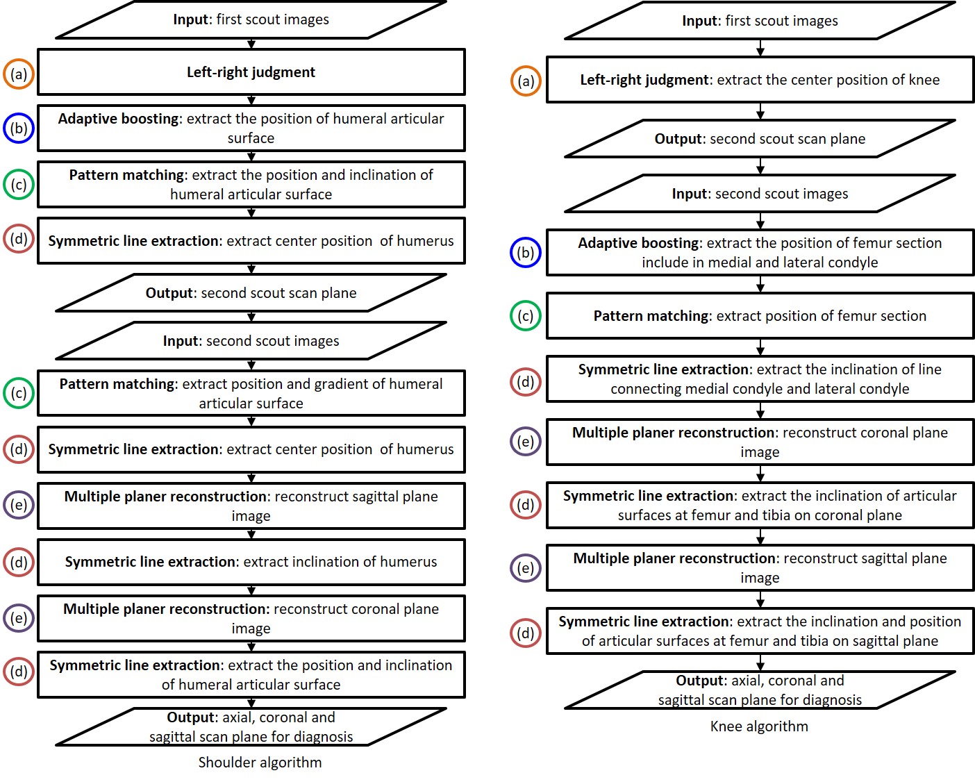
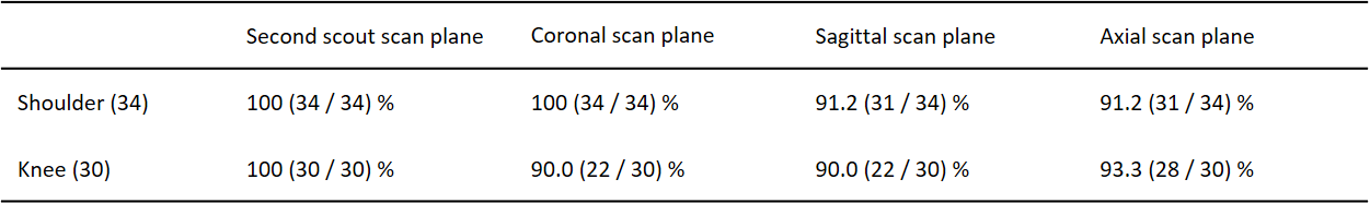
A flow chart of the proposed algorithms is shown in Figure 1. In this method, the scan planes are automatically prescribed using two sets of 2D scouting images. First scout images (axial images) are used to identify the left and right positions and extract scan planes for second scout imaging. Second scout images (axial, coronal, and sagittal images) are used to extract sagittal, coronal and axial scan planes for diagnosis. The feature extraction processes commonly used in the algorithms for knee and shoulder are as follows: (a) left-right judgment process by region growing method, (b) object extraction process by Adaptive boosting3, (c) object extraction process by pattern matching method, (d) symmetry line extraction process using correlation between left and right regions, and (e) multiple planar reconstruction process. The combination of each common process is modified in shoulder and knee algorithms. The shoulder algorithm extracts a line connecting the humerus and the scapula in the axial image, inclination of the humerus in the sagittal image, and inclination and position of the articular surfaces consisting of the humerus and scapula in the coronal image. The knee algorithm extracts a line connecting the medial and lateral condyle of the femur in the axial image, and inclination and position of the articular surfaces consisting of the femur and tibia in the coronal and sagittal images, respectively.For evaluation, scout imaging was performed on 34 shoulder images and 31 knee images using 1.5 T system. The study was approved by the ethics committee of Hitachi group headquarters. The accuracy evaluation was considered successful if the output of scan planes was within 25 mm of center position and ± 5 degrees of angle.
Results
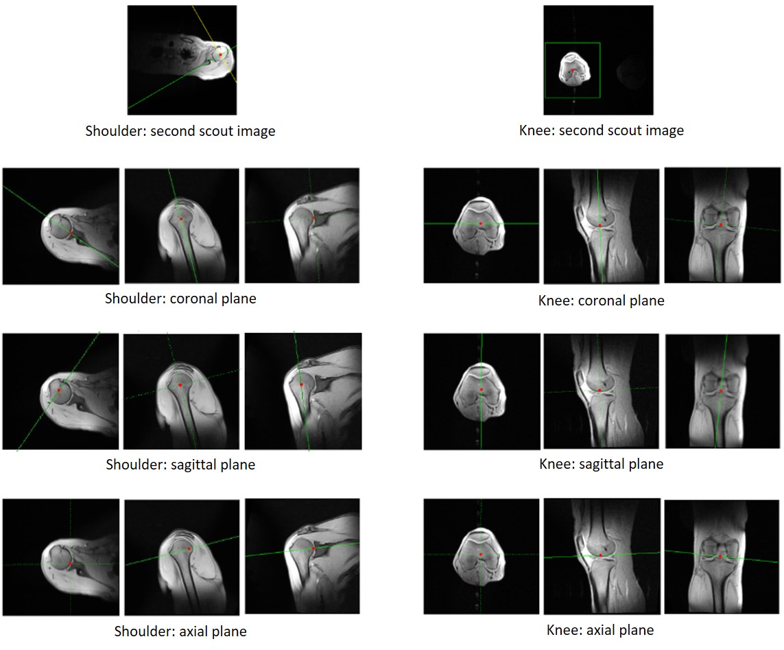
Figure 2 shows an example of automatically prescribed scan planes. In all cases, the automatically priscirbed scan planes did not deviate significantly. The results of evaluation are summarized in Table 1. The success rates of extracting the second scout scan plane using the first scout images were 100 % for shoulder and knee. The success rates of extracting coronal scan plane were 100 % for shoulder and 90.0 % for knee. The success rates of extracting sagittal scan plane were 91.2 % for shoulder and 90.0 % for knee. The success rates of extracting axial scan plane were 91.2 % for shoulder and 93.3 % for knee. Processing time was about 3 seconds, executing offline processing with CPU: Intel Core i7-6700k 4.00 GHz calculator.Discussion
Automated scan plane planning for shoulder and knee were proposed by modular algorithms development method. The results suggested that it is possible to realize automated scan plane planning for multiple examination parts by simply changing the combination of common processing. The reason for non-success case of the shoulder algorithm was poor extraction of the inclination and position of the articular surfaces consisting of the humerus and scapula in the coronal images by pattern matching process. Since there are individual differences in the tissue structure of the articular surfaces, the accuracy of the algorithm could be improved by using a template that takes deformations into account instead of a fixed shape template. The main reason for non-success case of the knee algorithm was poor extraction of the inclination of the medial and lateral condyle. This could be improved by optimizing the region where symmetry is calculated. The images used in this evaluation were only from healthy volunteers and adults, and the accuracy for disease cases and children has not been fully evaluated. In the future, clinical data and data from a wide range of age groups should be.Conclusion
We have proposed a modular algorithm developing method for automatic scan plane planning and developed the algorithms for shoulder and knee by changing the combination of common processes. It was suggested that this method may allow the automatic scan plane planning for more examination parts.Acknowledgements
No acknowledgement found.References
1. Yokosawa S, Taniguchi Y, Bito Y, et al. Automated scan plane planning for brain MRI using 2D scout images, Proceeding of ISMRM 18, 2010: 3136.
2. Yokosawa S, Noguchi Y, Sakuragi K, et al. Robust detection of anatomical landmark by combining adaptive boosting and active shape model for automated scan plane planning of spine MRI, Proceeding of ISMRM 27, 2019: 4817.
3. Freund Y, Schapire RE, A decision – theoretic generalization of on-line learning and an application to boosting, Journal of computer and system sciences 1887;55(1):119-139.
Figures