3704
Discrimination of IDH1 Genotype and 1p/19q Status in Glioma: A comparison study between Arterial Spin Labeling and Amide Proton Transfer imaging1Department of Magnetic Resonance, Lanzhou University Second Hospital, Lanzhou, China, 2Philips Healthcare, Xi'an, China
Synopsis
We investigated the association of amide proton transfer weighted imaging (APTw) and 3D arterial spin label (3D-pCASL) with IDH1 Genotype and 1p/19q Status in gliomas,then compared the two methods. All patients underwent MR examination, including APT and 3D-pCASL scan to get their values of the solid part. The results showed both APTw and 3D-pCASL had predictive values on IDH1 Genotype and 1p/19q Status in gliomas, and the performance of 3D-pCASL was better. We conclude that both APTw and 3D-pCASL can be used to predict the gene type of IDHin high-grade and 1p/19q co-deletion of gliomas before surgery, meanwhile 3D-pCASL is the better choice.
Introduction
Gliomas are the most common primary brain tumors. According to the WHO criteria 2016, molecular features have been incorporated into the classification of brain tumors. Isocitrate dehydrogenase (IDH) genotypes and status of chromosome arms 1p and 19q (1p/19q) has become important parts for subtyping[1]. However, it is not uncommon that lab tests lead to false-negative results when inadequate neoplastic cells from a biopsy procedure are mixed in with the normal cell background. It is necessary for researchers to find a noninvasive way to help diagnose and provide a comprehensive assessment. APTw imaging is a novel molecular imaging technique, which can noninvasively detect the exchange rate between amide protons and water-hydrogen ions[2]. In lesions, the number of mobile protons in proteins or peptides is usually varied at different stages and also shows difference in gliomas. 3D-pCASL, as a noninvasive perfusion magnetic resonance (MR) imaging technique without administration of exogenous contrast agent, that can qualitatively and quantitatively evaluate cerebral blood flow (CBF). In this study, we intend to investigate the merit in predicting IDH、1p/19q genotypes with APTw and 3D-pCASL in gliomas[3], providing the accurate gene information before surgery.Methods
This study recruited 30 patients from Lanzhou university Second Hospital. 13 patients were excluded, for their histopathological results couldn’t be obtained through surgical resection .Among the patients included, 10 cases were IDH mutant type and 7 cases were IDH wild type, 9 cases were 1p/19q codeletion type and 8 cases were 1p/19q undeletion type. Routine sequences like T1W enhancement, APT and 3D-pCASL were carried out on a 3T scanner (Ingenia CX, Philips Healthcare, the Netherlands) using a 32 channel head coil. 3D-pCASL imaging was obtained by using a three dimensional pseudo continuous pulse sequence. APT imaging was performed with 3D TSE-DIXON sequence. B0 corrected APT images were reconstructed automatically online by the software. 3D-pCASL and APT images were automatically coregistered to the FLAIR and post-contrast 3D-T1W images by performing a rigid transformation of the datasets. In order to accurately define the tumor borders. The region of maximal abnormality within the lesion volume (hotspot) was determined with a visual inspection on 3D-pCASL and APTw maps. Three separate ROIs were placed on the hotspot, avoiding intra-tumoral blood vessels, hamorrhage, cystic or necrotic regions, and the mean value for each parameter (APTw and 3D-pCASL) was recorded. The correlation of the four groups of data was tested by Spearman's rank correlation coefficient. Receiver operator characteristic (ROC) and area under curve (AUC) was performed to determine the diagnostic efficiency.Results
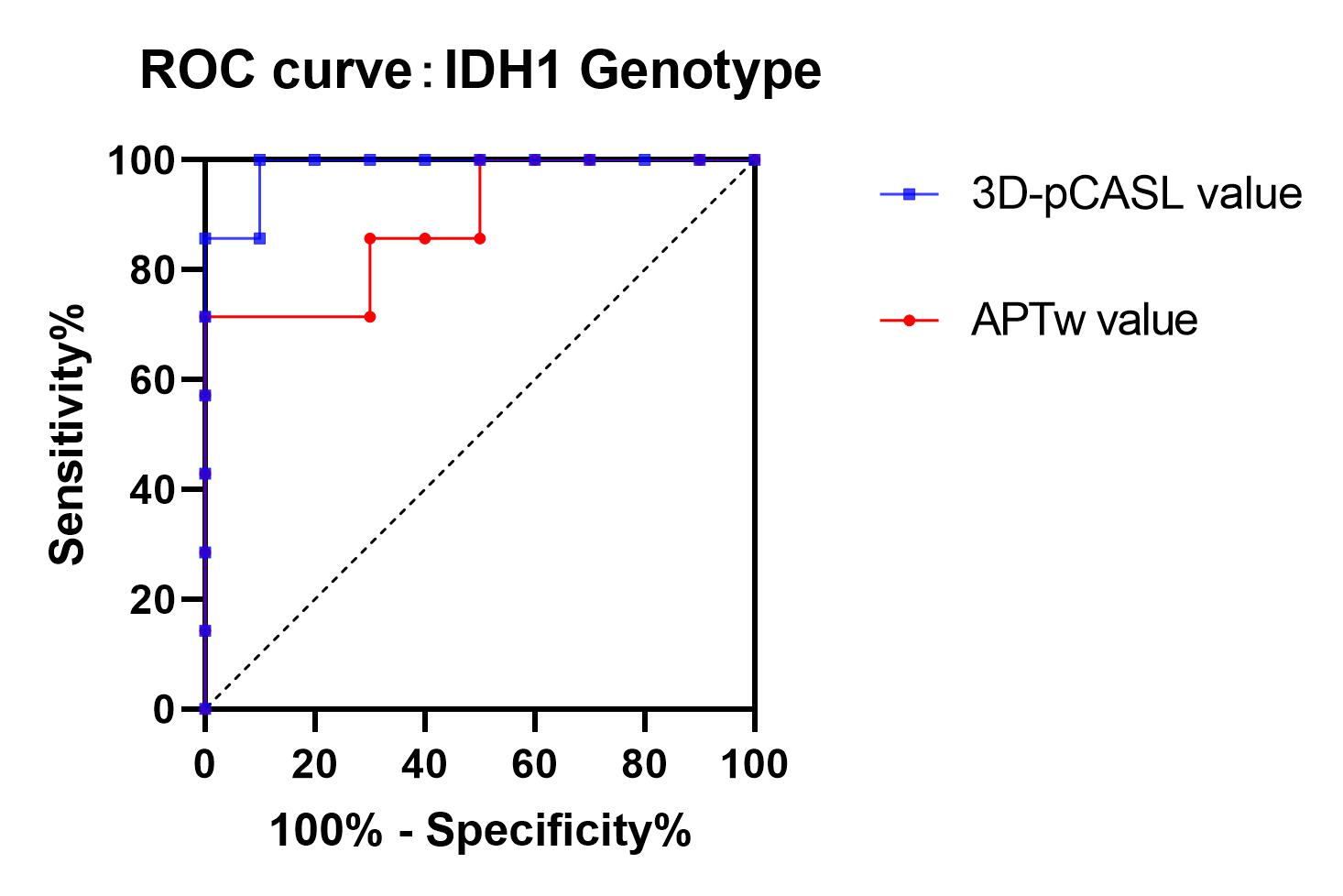
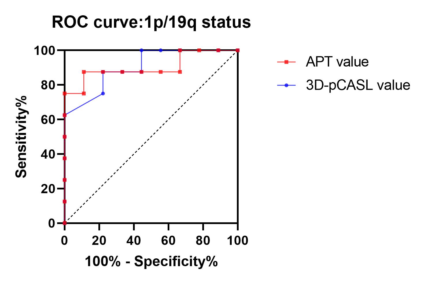
The ratio of IDH-1 wild type and 1p/19q undeletion increases with APTw value and 3D-pCASL value increasing(P<0.05). Sensitivities, Specificities and AUC of IDH1 genotype group (APTw,3D-pCASL) are 85.71% and 100.00%, 70.00% and 90.00%, 0.885 and 0.985. The same statistical results of 1p/19q status group (APTw, 3D-pCASL) are 87.50% and 87.50%, 77.78% and 88.89%, 0.903 and 0.903 (Table 1). The 3D-pCASL is better than APTw. When the tumor APT value and 3D-pCASL value was higher than 2.12% and 44.75 (mg/100ml/min), the patient was likely to be IDH wild type. When they are higher than 3.4% and 87.5(mg/100ml/min), the patient was likely to be 1p/19q undeletion.Discussion
Previous studies have shown that the presence or absence of IDH mutation and 1p/19q codeletion affects the prognosis of gliomas. The IDH1 enzymes are located in the cytosol/peroxisome, which has shown correlation with several important cellular reactions, including histone modifications, hypoxia sensing, and fatty acid metabolism.1p/19q is typically associated with mutations in IDH1. IDH mutation type and 1p/19q codeletion present more in high-grade glioma. The high-grade glioma accompanies with more cellular metabolism, including more protein and more perfusion.[4,5] Vasorin,a transmembrane protein, even can stimulate glioma growth and angiogenesis[6]. Zhou et al demonstrated that APTw can be used to detect brain tumors, predominantly on the basis of their higher protein/peptide content compared to brain tissue[7]. As Alberto Falk Delgado described[8], ASL can non-invasively map the entire tumor and quantify cerebral blood flow (CBF), a marker of angiogenesis and thus of tumor grade. As far as we know, there is no research compared the APTw and 3D-pCASL in predicting IDH1 Genotype and 1p/19q Status. The result of this study showed both APTw and 3D-pCASL can predict IDH1 Genotype and 1p/19q Statuswhich is consistent with previous studies. But 3D-pCASL had higher sensitivity and specificity than APTw. The possible reason may be the following points: 1) APTw value also is affected by PH environment. A research showed IDH-mutant tumors have significantly lower lactate concentrations[5], which may change PH environment resulting in inaccurate consequence. 2) Perfusion has positive correlation with vascular density, a directly maker of tumor grade. Cerebral blood flow (CBF) can be evaluated quantitatively by 3D-pCASL whether under physiological or pathological conditions. It is less affected by other factors. Therefore, 3D-pCASL has its unique advantages.Conclusion
The present study is the first analysis to compare APTw and 3D-pCASL on the predictive value of IDH1 genotype and 1p/19q status. Both APTw and 3D-pCASL have the predictive value of IDH1 genotype and 1p/19q status before surgery, the 3D-pCASL is better. It might be helpful for method choosing and clinical management .Acknowledgements
This research was supported by the Talent Innovation and Entrepreneurship Project of Lanzhou Chengguan District (Grant NO.2020RCCX0034).References
1. Eckel-Passow JE, Lachance DH, Molinaro AM. Glioma Groups Based on 1p/19q, IDH, and TERT Promoter Mutations in Tumors. N Engl J Med. 2015 Jun 25;372(26):2499-508.
2. Dou W, Lin CE, Ding H. Chemical exchange saturation transfer magnetic resonance imaging and its main and potential applications in pre-clinical and clinical studies. Quant Imaging Med Surg. 2019 Oct;9(10):1747-1766.
3. Wang N, Xie SY, Liu HM. Arterial Spin Labeling for Glioma Grade Discrimination: Correlations with IDH1 Genotype and 1p/19q Status. Transl Oncol. 2019 May;12(5):749-756.
4. Jiang S, Zou T. Predicting IDH mutation status in grade II gliomas using amide proton transfer-weighted (APTw) MRI. Magn Reson Med. 2017 Sep;78(3):1100-1109.
5. Wenger KJ, Steinbach JP, Bähr O. Lower Lactate Levels and Lower Intracellular pH in Patients with IDH-Mutant versus Wild-Type Gliomas. AJNR Am J Neuroradiol. 2020 Aug;41(8):1414-1422.
6. Liang W, Guo B, Ye J. Vasorin stimulates malignant progression and angiogenesis in glioma. Cancer Sci. 2019 Aug;110(8):2558-2572.
7. Zhou J, Lal B, Wilson DA. Amide proton transfer (APT) contrast for imaging of brain tumors. Magn Reson Med. 2003 Dec;50(6):1120-6.
8. Falk Delgado A, De Luca F, van Westen D. Arterial spin labeling MR imaging for differentiation between high- and low-grade glioma-a meta-analysis. Neuro Oncol. 2018 Oct 9;20(11):1450-1461.
Figures
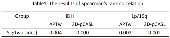
Table 1: Correlation analysis of APTw, 3D-pASL with IDH and 1p/19q