3691
Upright MR imaging of daily variations in pelvic organ position for assessment of pelvic organ prolapse1Magnetic Detection & Imaging, TechMed Centre, University of Twente, Enschede, Netherlands, 2Multi-Modality Medical Imaging, TechMed Centre, University of Twente, Enschede, Netherlands, 3Department of Gynecology, Ziekenhuisgroep Twente, Almelo/Hengelo, Netherlands
Synopsis
The choice of treatment for pelvic organ prolapse (POP) depends on determining its maximal extent but this might be subject to daily variations. This study uses upright MRI to determine if pelvic organs descend during the day and to make a comparison between women with and without symptomatic POP. For now, only women without POP were scanned three times during one day in supine and upright position. On average the cervix descended during the day and this effect was larger in upright position. A similar effect is expected in patients making the time of staging POP clinically relevant.
Introduction
Pelvic organ prolapse (POP) is a common problem in middle aged women, with a prevalence of symptomatic POP of 11.4%1. POP can be categorized in different stages, varying from stage 0 (no prolapse) to stage 4 ((almost) total eversion of the lower genital tract). POP stage is determined by physical examination of the maximal extent of POP, and is one of the factors that is associated with treatment choice. The circumstances under which POP staging must be performed are not standardized. However, pelvic organ position, the POP stage and thereby the treatment may depend on the circumstances during POP staging since they are not standardized.It is hypothesized that pelvic organ position is subject to variation during the day because the ligaments that lift the organs are viscoelastic. This means that the moment at which POP staging is performed may influence the treatment. Therefore, in this study daily variation of pelvic organ position will be investigated. Menopause and vaginal delivery are also associated with the elasticity of the uterosacral ligament and therefore the extent of POP2. To test if daily variation is present in POP, and if it correlates to menopause and vaginal delivery, all factors will be evaluated separately in this study.
MRI is a useful technique for the evaluation of pelvic organ position since it allows for a multicompartment assessment; it can render three-dimensional images of the bladder, uterus and rectum simultaneously. Furthermore, upright MRI allows visualization of the pelvic organs in different body positions3, which is important because previously it was proven that the extent of POP in upright patient position is significantly larger than in supine position4.
The aims of this study are:
- to assess the added value of upright MRI in evaluation of daily variation in pelvic organ position
- to evaluate the possible influence of daily variation on POP staging in symptomatic patients
- to compare differences in pelvic organ position during the day between symptomatic patients and women without symptomatic POP in different stages during life
Methods
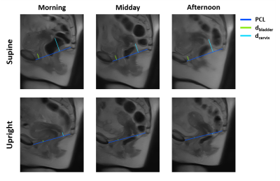
In this study 60 women will be included, divided in four groups: 1) nulliparous pre-menopausal women without symptomatic POP, 2) pre-menopausal women with ≥1 vaginal delivery without symptomatic POP, 3) post-menopausal women with ≥1 vaginal delivery without symptomatic POP, 4) post-menopausal women with ≥1 vaginal delivery and symptomatic POP. Due to the COVID-19 crisis, until now, only 4 women from group 2 were included. Therefore, for now only the first study aim could be investigated. However, the methods described below will also be applied to all subsequent women who will be included in this study.The women were scanned using a 0.25T MRI scanner (G-scan Brio, Esaote SpA, Italy). Three MRI scans in supine and upright position were performed during one day (morning, midday and afternoon). A sagittal multi-slice T2-weighted fast spin echo (FSE) (TE/TR: 25/3480 ms, flip angle: 90°, reconstructed resolution: 1.3 × 1.3 mm2, slice thickness: 5 mm, FOV: 340 x 340 mm2 number of slices: 11, echo train length: 10, echo spacing: 25 ms, total scan time: ≈2 min) was acquired. The positions of the bladder and the cervix were defined as their perpendicular distance to the pubococcygeal line (dbladder and dcervix respectively), which is commonly used (Figure 1). Positions above the line were indicated as negative and positions below the line as positive distances. Because only four women were included, the effect during the day was determined by calculating the median of the difference between the morning and afternoon measurements.
Results
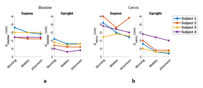
A small decrease in bladder position during the day was noticed, which was comparable between supine (median 0.1 cm) and upright position (median 0.2 cm) (Figure 2a). In upright position the cervix descended during the day in all subjects, while in supine 2 out of 4 subjects had comparable cervix positions (Figure 2b). The median differences between morning and afternoon in dcervix are 1.0 and 0.5 cm in upright and supine position respectively.Discussion
Although the study sample is very limited until now, we have demonstrated that in asymptomatic, pre-menopausal, parous women the cervix descends during the day, and that this effect is larger in upright position. All subjects need to be included to determine if there is a true effect of daily variation on bladder and cervix position and to statistically analyze differences between groups. Since symptomatic patients experience more symptoms towards the end of the day5, it is expected that the daily variation in patients will be at least as high as in asymptomatic controls. Because the difference between two POP stages can be as little as 2 cm, the stage may be underestimated when measured in the morning and thereby treatment selection can be suboptimal.Conclusion
Upright MRI delivers more insight in the effect the moment of the day has on the position of pelvic organs. In asymptomatic, pre-menopausal women with ≥1 vaginal delivery the cervix seems to descend during the day. When an effect of daily variation in pelvic organ position will be found in patients, clinicians can determine the ideal moment of the day to assess POP stage and thereby select the therapy for the patient.Acknowledgements
No acknowledgement found.References
1. Slieker-ten Hove MCP, Pool-Goudzwaard AL, Eijkemans MJC et al., Symptomatic pelvic organ prolapse and possible risk factors in a general population. Am J Obstet Gynecol. 2009;200(2):184.e1-184.e7.
2. Reay Jones NHJ, Healy JC, King LJ, Saini S, et al., Pelvic connective tissue resilience decreases with vaginal delivery, menopause and uterine prolapse. Br J Surg. 2003;90(4):466-472.
3. Williams HG, Cooper A., Hodgen L et al., Upright pelvimetry using MRI for the prediction of birth associated cephalo-pelvic disproportion following induction of labour. Proc. Intl. Soc. Mag. Reson. Med. 28 (2020)
4. Grob ATM, Heuvel J olde, Futterer JJ, et al., Underestimation of pelvic organ prolapse in the supine straining position, based on magnetic resonance imaging findings. Int Urogynecol J. 2019;30(11):1939-1944.
5. Sung VW, Clark MA, Sokol ER, et al., Variability of current symptoms in women with pelvic organ prolapse. Int Urogynecol J. 2007;18(7):787-798.
Figures

