3688
The value of R2* in evaluating aggressiveness of ovarian tumors.1The First Affiliated Hospital of Dalian Medical University, Dalian, China
Synopsis
The aggressiveness level of ovarian tumor is of great importance for clinical treatment and prognosis. The purpose of this study is to explore the value of R2 * in evaluating the aggressiveness of ovarian tumors. The preliminary results showed that the R2 * value of malignant tumors was significantly higher than benign and borderline tumors. R2* is potentially a promising and valuable non-invasive method in evaluating the aggressiveness of ovarian tumors.
Introduction
Ovarian tumors can be classified into benign,borderline and malignant tumors depending on the tumor aggressiveness. Benign tumors could be treated with fertility sparing surgery,1 while the main treatment of malignant tumors is comprehensive staging surgery and adjuvant chemotherapy.2 Thus accurate preoperative diagnosis is of great significance in deciding the optimal surgical procedure and improving the life quality of patients. R2* is transverse relaxation rate that is obtained via gradient reunion at different times. The R2* value is directly related to the tissue deoxygenated hemoglobin concentration and can be used to quantitatively assess changes of oxygen content in local tissue. 3The purpose of this study was to explore the performance of R2* in classification of ovarian tumor aggressiveness.Methods
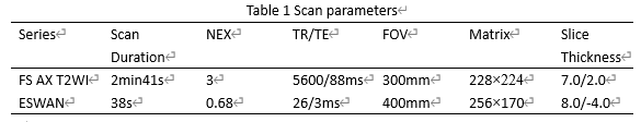
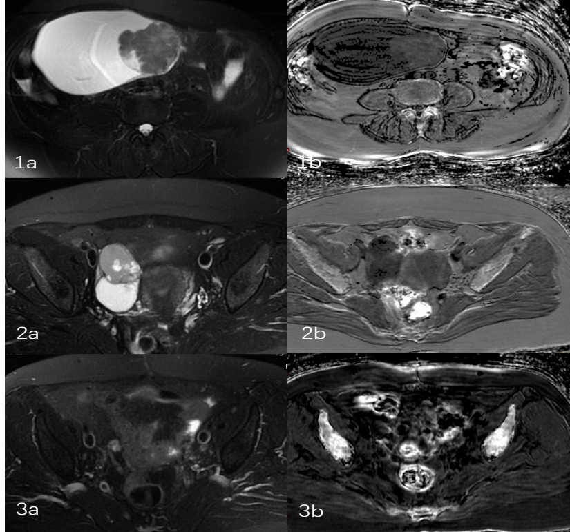
99 patients with ovarian tumors who were underwent T2-weighted imaging (T2WI) and enhanced T2 star-weighted angiography (ESWAN) scans on a 1.5 T MR scanner (Signa HDxt, GE Medical Systems, USA) were involved in this study. Detailed scan parameters were listed in Table 1. All patients were divided into benign tumors group (group 1, n=45. mean age: 47.8±20.2 years, range: 11-81 years), borderline tumors group (group 2, n=12. mean age: 48.3±17.0 years, range: 16-83 years) and malignant tumors group (group 3, n=42. mean age: 53.4±14.7 years, range: 8-76 years). 1,2 and 3,4 groups were subsequently combined into group 4 (benign+borderline tumors) and group 5 (malignant+borderline tumors), respectively. The original axial digital images from the ESWAN sequence were transmitted to the ADW 4.6 workstation. Functool software was used to perform post-processing to obtain R2* images. With reference to fusion of T2WI and R2* images, the regions of interest (ROIs) were manually drawn on lesions of three slices (including the slice covering the largest dimension of tumor and its adjacent upper and lower slices) (Figure 1). The average R2* values were calculated to minimize measurement bias. All statistic analyses were analyzed by SPSS 26.0 software. The comparisons among the five ovarian tumor groups were performed using the Mann–Whitney U test with Bonferroni correction. The receiver operating characteristic (ROC) curves were used to examine the diagnostic performance characteristics of R2* value for ovarian tumors with different levels of aggressiveness.Results
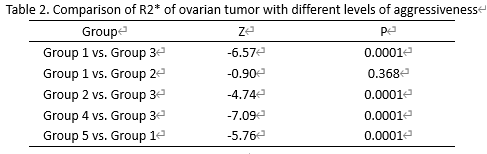
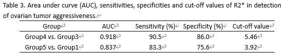
R2* value of group 3 was significantly higher than that of group 1 (10.70 ±4.73 vs. 3.60±4.48s-1, p < 0.0001) and 2 (10.70±4.73 vs. 3.25 ±1.85s-1, p<0.0001). No significant difference of R2* value was observed between 1 and 2 groups. R2* value of group 4 was significantly lower than that of group 3 (3.53±4.06 vs. 10.70±4.73s-1, p<0.0001). R2* value of group 5 was significantly higher than that of group 1 (9.05±5.27 vs. 3.60±4.48s-1, p<0.0001). (Table 2). The AUC value for the ROC analyses of R2* value for differentiation between group 4 and group 3 was 0.918. The sensitivity and specificity were 90.5% and 86%, respectively, with a cut-off value of 5.46. The AUC value for the ROC analyses of R2* value for differentiation between group 5 and group 1 was 0.837. The sensitivity and specificity were 83.3% and 75.6%, respectively, with a cut-off value of 3.92.(Table 3)Discussion and Conclusion
Our study indicated that the R2* value of malignant tumors was higher than benign and borderline tumors. Compared with benign and borderline tumors, malignant tumors are more invasive and have higher proliferation rate, which increases oxygen consumption. The lower oxygen content and increase in deoxygenated hemoglobin lead to a high R2* value. When the R2* value of an ovarian tumor is less than 3.92, it can be diagnosed as benign tumor. When the R2* value of an ovarian tumor is greater than 5.46, it can be diagnosed as malignant tumor. When the R2* value of an ovarian tumor is between 3.92 and 5.46, its aggressiveness level is uncertain This study suggested that R2* could be used to effectively determine the aggressiveness level of ovarian tumor and provide a basis for clinical treatment.Acknowledgements
No acknowledgement found.References
1. Wang Ming, Wu Yumei. Fertility preservation techniques for gynecological benign tumor surgery. Chinese Family Planning and Obstetrics and Gynecology,2020,12(10):21-24.
2. Wang Xiaoni, Wang Qian, Tian Xiaojuan, Suo Yuping.Current status and progress in the treatment of ovarian cancer. Anti-tumor Pharmacy,2020,10(03):264-268+286.
3. Bai Xiaoxi, Zhang Liou, Huang Liping. Progress and clinical application of magnetic resonance ESWAN imaging technique. Modern oncology, 2019,27 (09): 1621-1624.
Figures