3687
A Pilot Evaluation of Intravoxel Incoherent Motion (IVIM) in Characteristics and Diagnosis of Ovarian cancer of p53 status
Yulin Chen1, Ye Li1, li Liu1, Xinliu He1, Xulun Lu1, and Ailian Liu1
1The First Affiliated Hospital of Dalian Medical University, Dalian, Dalian, China
1The First Affiliated Hospital of Dalian Medical University, Dalian, Dalian, China
Synopsis
The identification and treatment of ovarian cancer(OC) has always been a problem that plagues us[1]. This worked aimed at exploring the value of Intravoxel incoherent motion (IVIM) quantitative parameters for diagnosis of P53 positive OC. In contrast, P53 positive and negative OC have large differences in stand ADC values in IVIM.
Introduction
Ovarian cancer (OC) is a common tumor, accounting for 23% of female reproductive malignancies, and its incidence is increasing year by year[1]. Because ovarian cancer has no obvious clinical symptoms in the early stage, even if there are symptoms, there is no specificity, and the role of screening is very limited. Moreover, the development is relatively fast, and many patients have advanced ovarian cancer at the time of treatment. The expression of mutant p53 or loss of p53 allows ovarian cancer cells to continue to proliferate. In various genetic related diseases and malignant tumors, there are deletions or mutations in the P53 gene, which are related to the growth and development of malignant tumors. P53 plays an important role in tumor formation. This study aims to explore the relationship between IVIM quantitative parameters and P53 protein in OC, In this study, we used stand ADC, D, D* and f value of IVIM for analysis to explore the difference between P53 positive and negative OCMethods
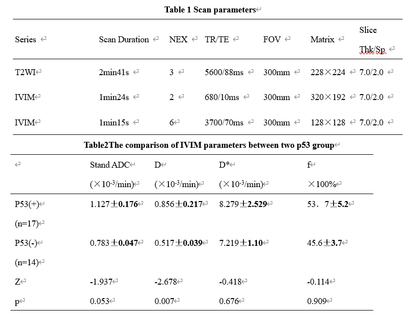
A total of 32 patients of ovarian cancer confirmed by surgery and pathology from 2015 to 2020 were retrospectively collected. Including 18 P53-positive patients (age:51.71±11.08 years old; range:(35,74) and.14 negative patients (age 60.82±8.65years old; range:(47,76) ). All patients underwent abdominal MR examinations (Signa HDxt, GE Medical Systems, USA) included T2WI, IVIM and LAVA(Scan parameters show in table 1). The original axial digital images from the IVIM sequence were transmitted to the GE SDC-ADW 4.6 workstation (Sun Microsystems, Santa Clara, Calif) and the post-processing was performed by Functool software. Referring to the anatomical location of lesion obtained on T2-weighted or DWI images, The stand ADC, D, D* and f maps were automatically constructed, and were reviewed by two observers who were blinded to clinical information and histopathologic results with 10 and 15 years of experience in pelvic imaging, respectively. The manual regions of interest (ROIs) were drawn along the edge of tumors on the slice with maximal solid area (we choose the solid in tumors), according to fatsuppression T2WI and T1WI (Fig. 1). The measurement was repeated for three times, and the averages of three measurements were calculated. The stand ADC, D, D* and f were recorded. Intra-group correlation coefficient (ICC) was used to test the measurement consistency between the two observers. Difference between above values and the expression of P53 in OC was compared by Mann-Whitney U test. Receiver operating characteristic (ROC) analysis was performed to evaluate diagnostic performance.Results
The stand ADC value of the positive group was significantly higher than that of negative group (p <0.05, Table 2). The ROC curve of the stand value in the mutant and wild-type groups showed an AUC value of 0.723 (Figure 1). When the threshold of stand ADC value is set to 0.999×10⁻³/min, the corresponding diagnostic sensitivity and specificity will be 58.8% and 92.9%.Discussion and Conclusion
p53 is a tumor suppressor protein. When p53 becomes mutated, it loses its function, resulting in abnormal cell proliferation and tumor progression[2]. The shrunken extracellular space due to abnormal proliferation of cancer cells and following incremental local cell density in malignant tumor tissues generates more severe diffusion limitation in malignant lesions than in benign lesions[3]. In the present study, we proposed that stand ADC value could be a promising imaging biomarker in evaluation of P53 expression.Acknowledgements
No acknowledgement found.References
[1]Al-AlemL,CurryJ.Ovarian cancer: involvement of the martrixmetall oproteinases(J).Reproduction,2015,150(2):55-64[2]Mathumai Kanapathipillai. Treating p53 Mutant Aggregation-Associated Cancer. Cancers (Basel). 2018 May 23;10(6):154.[3] Long L, Zhang H, He X, Zhou J, Guo D, Liu X. Quantitative evaluation of intravoxel incoherent motion diffusion-weighted imaging (IVIM) for differential diagnosis and grading prediction of benign and malignant breast lesions. Medicine (Baltimore). 2018 Jun;97(26):e11109.Figures
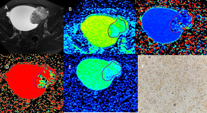
Figure 1 A 63 year-old woman with left side ovarian cancer. A T2WI shows a low signal lesion in left pelvis .B The stand ADC value is 1.09×10⁻³mm2/s. C The D value is 0.791×10⁻³mm2/s. D The D* value is 4.35×10⁻³mm2/s. E The f value is 0.621. F The p53 picture of this lesion.
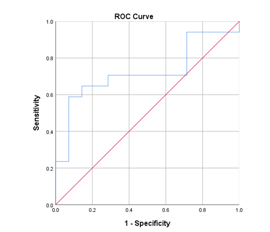
FIG1
Table