3686
Diffusion Properties of Ovaries in Pre-Menopausal Women Using a Bi-Exponential Model1Radiology, University of California San Diego, La Jolla, CA, United States, 2School of Medicine, University of California San Diego, La Jolla, CA, United States, 3University of California San Diego, La Jolla, CA, United States
Synopsis
Ovarian cancer has a high mortality rate as it is commonly diagnosed in advanced stages. Diffusion weighted imaging (DWI) has shown great promise in detecting cancer in multiple other organs. However, the diffusion characteristics of premenopausal ovaries are difficult to differentiate from malignancies with standard diffusion techniques. In this work we characterized the diffusion properties of ovaries in a group of young women to inform the acquisition and post-processing of DWI to improve ovarian cancer diagnosis.
Introduction
Ovarian cancer has a high mortality rate as it is commonly diagnosed in advanced stages.1 Diffusion weighted imaging (DWI) has shown great promise in detecting cancer in multiple other organs. Unfortunately, the differentiation between benign and malignant ovarian tissues represents a challenge with standard DWI techniques as ovaries present high intensity MR signal at high b-values. Although previous studies have suggested that the diffusion-weighted signal decay of ovarian lesions is biexponential, data describing the signal of healthy ovaries are lacking. Characterization of the diffusion properties of healthy ovaries would allow for tailoring of DWI data acquisition and post-processing to improve the detection of ovarian cancer. Thus, the purpose of this work was to evaluate the diffusion properties of ovaries throughout the menstrual cycle using a biexponential model.Methods
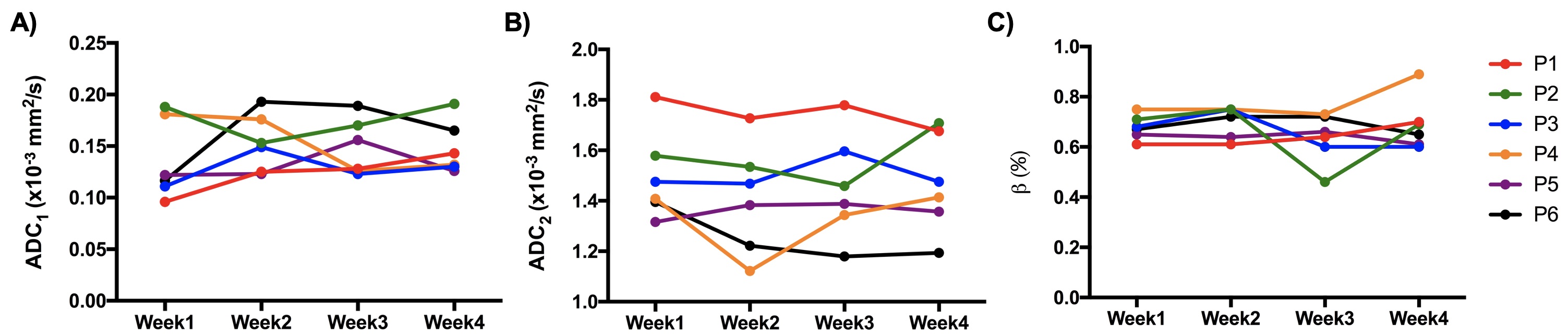
Six women (26±6 years old) with regular 28-day menstrual cycle underwent pelvic MRI in a 3T scanner using a 32-channel body array over four consecutive weeks. Imaging parameters: T2 FSE— TE/TR=100/7690ms, FA=140°, FOV=200×200mm2, voxel size=0.4×0.4×3mm3; Multi-shell DWI— TE/TR=65/8075ms, FOV=160×160mm2, voxel size=1.56×1.56×3mm3, 6 diffusion directions were probed for b-values 0,125, 250, 375, and 750 s/mm2, and 12 directions for b=1000 and 2000s/mm2. Two b=0 s/mm2 volumes were acquired in positive and negative phase-encoding (PE) trajectory polarities to correct B0-inhomogeneity induced distortions using the reverse polarity gradient (RPG) method.3 All analyses were performed in MATLAB. Diffusion directions for a determined b-value were averaged before processing (assuming isotropic diffusion). Regions of interest (ROIs) in the ovaries were manually drawn on distortion corrected B0 images. Here, the diffusion-weighted signal was modeled as the linear combination of two exponential decays: $$S_i(b) = S_0[(1-\beta)e^{-b ADC_1}+\beta e^{-bADC_2}]$$ , where β is the weighting coefficient of each term, b are the b-values in s/mm2, and ADC1 and ADC2 are the ADCs of each exponential component (ADC1<ADC2) in mm2/s. We hypothesized that the diffusion characteristics of ovaries vary throughout the menstrual cycle as ovarian follicles prepare for ovulation (between weeks 2 and 3) and involute before the menstrual cycle (week 4). One-way repeated measures analysis of variance (ANOVA) was used to test for differences in diffusion characteristics throughout the menstrual cycle.Results
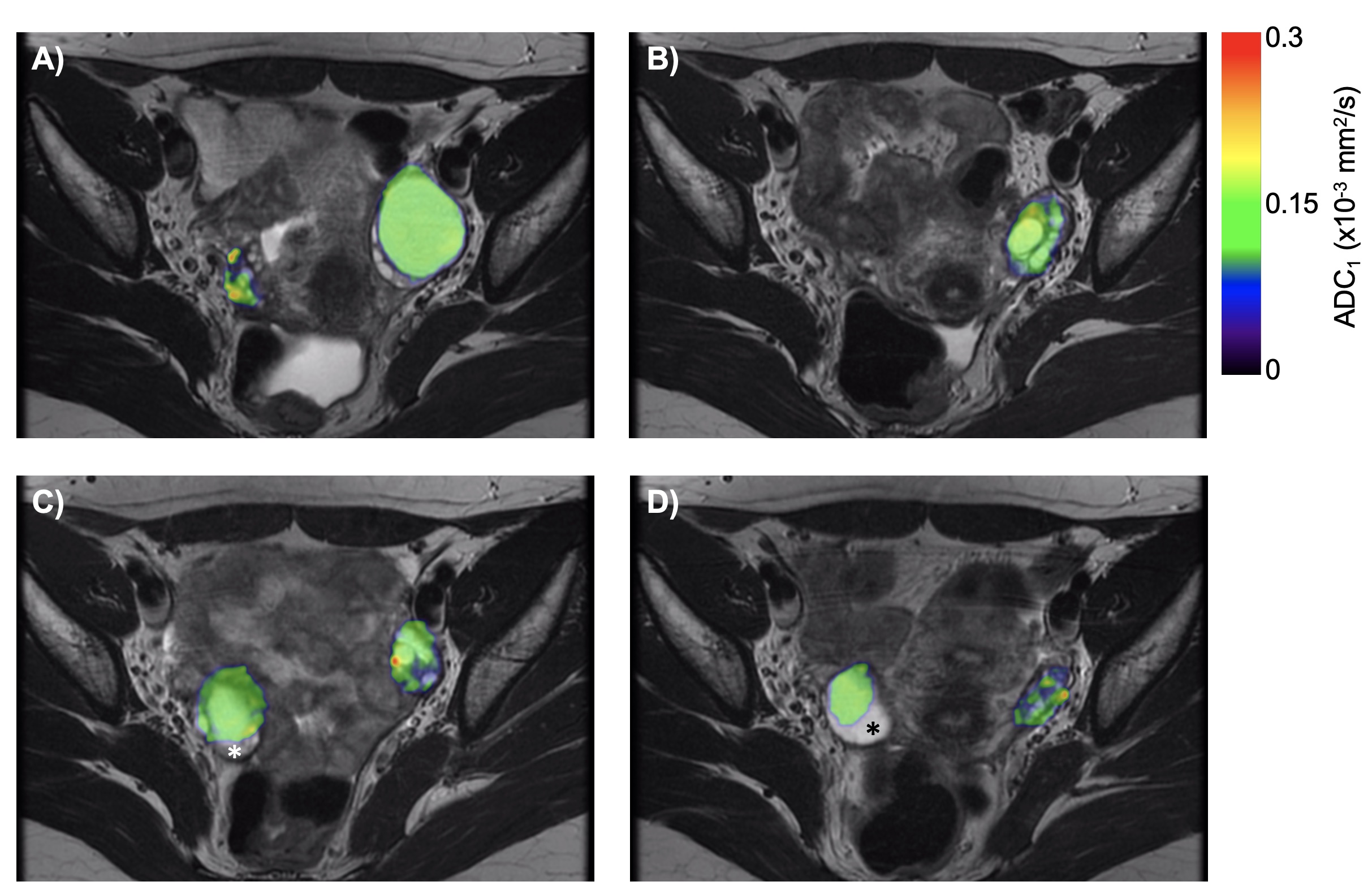
No differences in the diffusion properties of the ovaries were observed throughout the menstrual cycle. Representative ADC1 maps overlaid on anatomical images are shown in Fig. 1. Estimated average ADC1, ADC2 and values were 0.15±0.03×10⁻⁵mm2/s, 1.45±0.19×10⁻³ mm2/s and 0.68±0.08, respectively (Fig. 2).Discussion and Conclusions
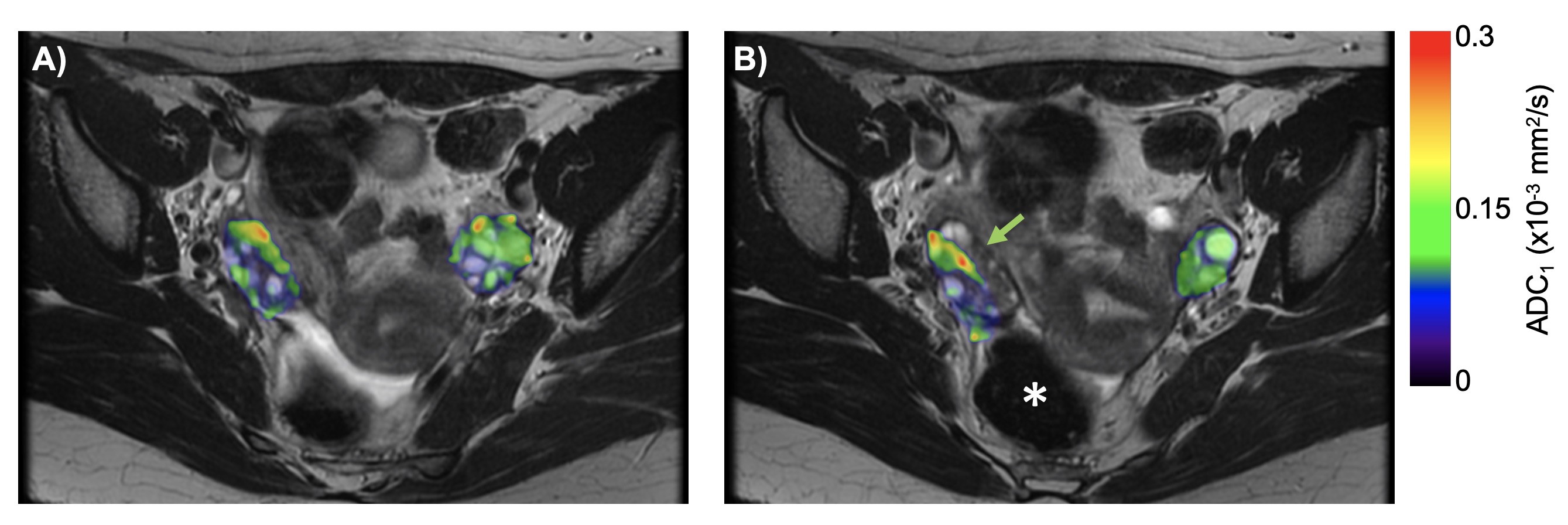
In this ongoing study, we report preliminary results of the diffusion properties of healthy ovaries throughout the menstrual cycle using a biexponential model. Natural variation in the diffusion properties of ovaries were expected throughout the menstrual cycle. Although this was not observed, it may be explained by the inclusion of both ovaries despite the presence of a single dominant follicle per cycle. Additional factors that may conceal variation in the diffusion signal throughout the menstrual cycle may be related to the geometric distortions in the pelvic region. The performance of the distortion correction algorithm varies across the imaging volume as bowel gas distribution is heterogeneous. In cases when the discrepancy between anatomical and DW images was pronounced, care was taken to avoid the inclusion of such areas in ROIs. As a result, in some cases it was not possible to achieve complete ovary volume in this analysis (Fig. 3). The estimated ADC values are lower than those previously reported for ovaries over the menstrual cycle.3 Discrepancies may be attributed to imaging parameters (maximum b-value) and more importantly, to the model describing the diffusion signal (mono- versus bi-exponential). The appropriate characterization of the diffusion-weighted signal from both benign and malignant ovarian tissues is key to tailor DWI studies for the early detection of ovarian cancer. Similarly, multi-exponential DWI methods may improve the conspicuity of ovarian tumors. Future work will focus on characterizing the diffusion properties of ovarian follicles excluding parenchymal tissue, and to evaluate the use of advanced DWI methods in the female pelvis for ovarian cancer diagnosis.Acknowledgements
GE HealthcareReferences
1. U.S. Cancer Statistics Working Group. United States Cancer Statistics: 1999–2014 Incidence and Mortality Web-based Report. Atlanta (GA): Department of Health and Human Services, Centers for Disease Control and Prevention, and National Cancer Institute. 2017.
2. Holland D, Kuperman JM, Dale AM. Efficient correction of inhomogeneous static magnetic field-induced distortion in Echo Planar Imaging. Neuroimage. 2010;50(1):175-183.
3. Morisawa N, Kido A, Koyama T, et al. Changes of the normal ovary during menstrual cycle in reproductive age on the diffusion-weighted image. J Comput Assist Tomogr. 2012;36(3):319-322.
Figures