Yuan Wei1, Shifeng Tian1, and Ailian Liu1
1Department of Radiology, the First Affiliated Hospital of Dalian Medical University, China, Dalian City, China, China
Synopsis
Diffusion tensor imaging (DTI) is a functional imaging technology improved and developed on the basis of DWI technology. It reflects the changes of free diffusion rate and direction of water molecules caused by different structures in living tissues. It can obtain multiple quantitative parameters through post-processing. There is no report on the evaluation of endometrial carcinoma (EC) microsatellite instability (MSI) status by DTI quantitative parameters. Results shown that multiple quantitative parameters of DTI were different between MSI and microsatellite stability (MSS) of EC, therefore, it is feasible to use DTI for predicting MSI of EC.
INTRODUCTION
Microsatellite instability (MSI) can be used as an indicator to screen for Lynch syndrome (LS) related endometrial carcinoma (EC), evaluate the efficacy of immunosuppressive therapy and evaluate the prognosis[1]. Diffusion tensor imaging (DTI) can detect the diffusion rate and direction of water molecules in voxels. It can describe the diffusion motion of water molecules more accurately and provide multiple quantitative parameters. The application of DTI in EC has been reported, which mainly focuses on the quantitative evaluation of EC myometrial infiltration, pathological classification and grading by DTI parameters[2,3]. The purpose of this study was to explore the value of quantitative parameters of DTI in predicting MSI of EC.METHODS
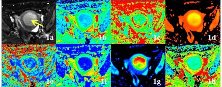
A retrospective analysis was performed on the data of 37 patients of EC confirmed by surgical pathology, which included 12 cases of MSI and 25 cases of microsatellite stability (MSS). The preoperative MR scanning sequence included DWI and DTI (1.5T, SignaHDxt, GE Medical Systems, USA). The apparent diffusion coefficient (ADC) values of DWI, the average diffusion coefficient (DC avg), the fractional anisotropy (FA), the isotropy (Iso), the volume ratio anisotropy (VRA), the exponential attenuation (Exat) and the T2-weighted trace (T2-WT) values of DTI were measured (Fig 1). The consistency of the measurement results was tested, the correlation between ADC and DC avg values of the two groups was analyzed, the differences of various parameter values between the two groups were compared, and the efficacy of prediction of MSI with different parameters was evaluated and compared.RESULTS
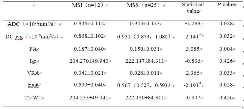
The data consistency of each group were good (ICC> 0.75). The ADC, DC avg values of MSI group were significantly lower than that of MSS group, the FA, VRA, Exat values of MSI group were significantly higher than that of MSS group(P<0.05). There was no statistical difference in Iso and T2-WT values between the two groups (P>0.05) (Table 1). The values of ADC and DC avg in MSI group and MSS group were highly correlated (r=0.906, 0.868, P<0.001). The AUC of EC MSI predicted by ADC, DC avg, FA, VRA and Exat values were 0.698, 0.720, 0.783, 0.743 and 0.725, respectively. There was no statistical difference among each AUC (P>0.05).DISCUSSION AND CONCLUSIONS
MSI related EC often accounted for a higher proportion, and more lymphocytes infiltrated tumor tissue. Therefore, MSI related EC tumor cell proliferation activity and ability is higher than MSS related EC, cell arrangement is more compact, extracellular space is reduced, water molecule diffusion is more restricted, which can lead to the decrease of DC avg value and the increase of Exat value. The tumor cells of MSI related EC are closely arranged, which may cause water molecules to spread along certain cell spaces and move more directionally, therefore, FA value and VRA value of MSI related EC increased. DTI can provide several quantitative parameters for predicting EC MSI, among which DC avg value is highly correlated with ADC value of DWI sequence.Acknowledgements
No acknowledgement.References
[1] Petrelli F, Ghidini M, Cabiddu M, et al. Microsatellite Instability and Survival in Stage II Colorectal Cancer: A Systematic Review and Meta-analysis. Anticancer Res, 2019, 39(12):6431-6441.
[2] Yamada I, Wakana K, Kobayashi D, et al. Endometrial carcinoma: Evaluation using diffusion-tensor imaging and its correlation with histopathologic findings. J Magn Reson Imaging, 2019, 50(1):250-260.
[3] Zhang L, Liu A, Zhang T, et al. Use of diffusion tensor imaging in assessing superficial myometrial invasion by endometrial carcinoma: a preliminary study. Acta Radiol, 2015, 56(10):1273-1280.