3682
Prediction of pretherapeutic differentiation degree of cervical cancer using DCE-MRI radiomics model1Radiology Department, Anhui Provincial Cancer Hospital, Hefei, China, 2Radiology department, Anhui Provincial Cancer Hospital, Hefei, China
Synopsis
The aim of the study was to explore the value of radiomics features based on DCE-MRI in Predicting the differentiation degree of cervical cancer before treatment. 97 patients pathologically confirmed cervical cancer from March 2017 to December 2018 were enrolled in this study. The conclusion of radiomics features based on DCE-MRI have good repeatability and are of high value for predicting pre-treatment differentiation of cervical cancer was obtained via the present study.
Objective
To explore the value of radiomics features based on DCE-MRI in Predicting the differentiation degree of cervical cancer before treatment.Methods
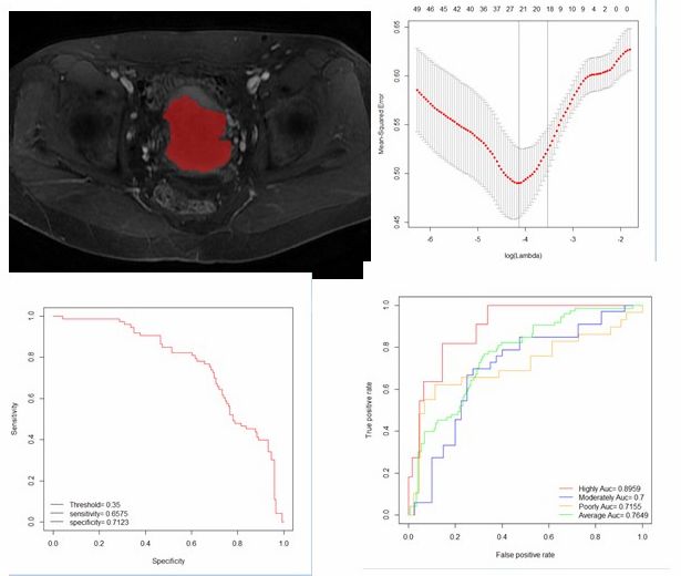
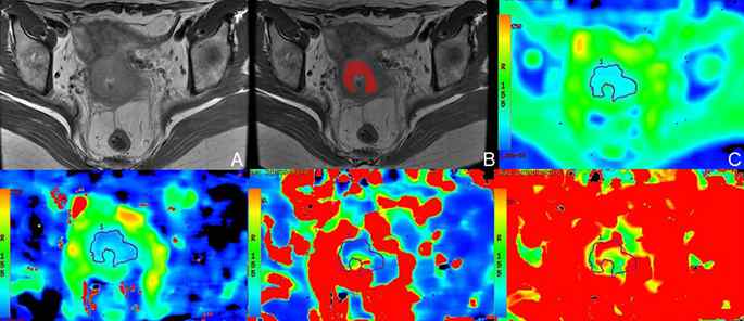
97 patients pathologically confirmed cervical cancer from March 2017 to December 2018 were analyzed retrospectively.54 cases were selected as training group,including 9 cases with high differentiation,20 cases with moderate differentiation, and 25 cases with poor differentiation.The other 43 patients were included in the validation group,including 7 with high differentiation, 17 with moderate differentiation, and 19 with poor differentiation. Routine MRI and DCE-MRI scans were underwent by all patients, and the largest level measurement of the penultimate phase in the original image of DCE-MRI was selected..ITK-SNAP and R language software were used for radiomics features extraction.The LASSO regression analysis was used for features selection.Logistic regression was performed to develop models and validation.The prediction performances were evaluated with ROC analysis and AUC curve.Results
A total of 945 features were obtained and reduced to six features as the most important discriminators for differentiation degree of cervical cancer.The prediction model was developed with maximum2DDiameterRow,glcm-ClusterShade,glszm-GrayLevelVariance,wavelet.LHL-Texture-ngtdm-Busyness,wavelet.HLL-FisrtOrder-Skewness and wavelet.LLL-FisrtOrder-Kurtosis.The radiomics features of the training group were tested by the verification group. The specificity of the specific-sensitivity curve obtained by logistic regression was 71.2%, the sensitivity was 65.8%, the positive predictive value was 0.762, and the negative predictive value was 0.684. The obtained characteristics were verified by the high, middle and low differentiation groups of the validation group. The area under the ROC was 0.896, 0.700, 0.716, the average AUC was 0.765, and the specificity was 86.4%, 69.2%, 76.1% respectively , sensitivity is 68.6%, 79.5%, 81.2%.Conclusion
Radiomics features based on DCE-MRI have good repeatability and are of high value for predicting pre-treatment differentiation of cervical cancer.Acknowledgements
No acknowledgement found.References
1. Thomassinnaggara I, Siles P, Balvay D, et al. MR perfusion for pelvic female imaging.[J]. Diagnostic & Interventional Imaging, 2013, 94(12):1291-1298.
2. Zhou Y, Liu J, Liu C, et al. Intravoxel incoherent motion diffusion weighted MRI of cervical cancer - Correlated with tumor differentiation and perfusion[J]. Magnetic Resonance Imaging, 2016, 34(8):1050-1056.
3. Han K, Croke J, Foltz W, et al. A prospective study of DWI, DCE-MRI and FDG PET imaging for target delineation in brachytherapy for cervical cancer[J]. Radiotherapy & Oncology, 2016, 120(3):519-525.
4. Lee E Y, Yu X, Chu M M, et al. Perfusion and diffusion characteristics of cervical cancer based on intraxovel incoherent motion MR imaging-a pilot study[J]. European Radiology, 2014, 24(7):1506-1513.
5. Kim J H, Kim C K, Park B K, et al. Dynamic contrast-enhanced 3-T MR imaging in cervical cancer before and after concurrent chemoradiotherapy[J]. International Journal of Medical Radiology, 2012, 22(11):2533.
6. Becker A S, Perucho J A, Wurnig M C, et al. Assessment of Cervical Cancer with a Parameter-Free Intravoxel Incoherent Motion Imaging Algorithm[J]. Korean Journal of Radiology, 2017, 18(3):510-518.
Figures