3681
Predictive Ki-67 proliferation index of cervical squamous cell carcinoma based on IVIM-DWI combined with texture features
Cuiping Li1 and Jiangning Dong2
1Radiology Department, The First Affiliated Hospital of USTC, Division of Life Sciences and Medicine, University of Science, Hefei, China, 2The First Affiliated Hospital of USTC, Division of Life Sciences and Medicine, University of Science and Technology of China, Hefei, China
1Radiology Department, The First Affiliated Hospital of USTC, Division of Life Sciences and Medicine, University of Science, Hefei, China, 2The First Affiliated Hospital of USTC, Division of Life Sciences and Medicine, University of Science and Technology of China, Hefei, China
Synopsis
As the most common histological type in cervical cancer, cervical squamous cell carcinoma (CSCC) mainly affects young women, and most of whom are diagnosed between 35-50 years old. In recent years, studies on tumor biomarkers have continuously increased, Ki-67 was considered as a kind of nuclear protein related to cell proliferation, some previous studies have shown that Ki-67 proliferation index (PI) is positively correlated with tumor size, invasion, cancer stage and patient survival. In our study work, we investigated the prediction of Ki-67 PI through combining intravoxel incoherent motion diffusion weighted imaging (IVIM-DWI) with texture analysis (TA) for CSCC patients.
Abstract
Purpose: This study aims to determine whether IVIM-DWI combined with texture features based on preoperative IVIM-DWI could be used to predict the Ki-67 PI, which is a widely used cell proliferation biomarker in CSCC. Methods: A total of 70 patients were included. Among these patients, 16 patients were divided into the Ki-67 PI<50% group and 54 patients were divided into the Ki-67 PI≥50% group based on the retrospective surgical evaluation. All patients were examined using a 3.0 T MRI unit with one standard protocol, including an IVIM-DWI sequence with 10 b-values (0-1,500 sec/mm2). The maximum level of CSCC with a b value of 800 sec/mm2 was selected. The parameters (diffusion coefficient [D], microvascular volume fraction [f], and pseudodiffusion coefficient [D*]) were calculated with the ADW 4.6 workstation, and the texture features based on IVIM-DWI were measured using the GE AK quantitative texture analysis software. The texture features included the first order, GLCM, GLSZM, GLRLM, and wavelet transform features. The differences in IVIM-DWI parameters and texture features between the two groups were compared, and the ROC curve was performed for parameters with group differences, and in combination. Results: The D value in the Ki-67 PI≥50% group was lower than that in the Ki-67 PI<50% group (P<0.05). A total of 1,050 texture features were obtained using the AK software. Through univariate logistic regression, mPMR feature selection and multivariate logistic regression, three texture features were obtained: wavelet_HHL_GLRLM_ LRHGLE, lbp_3D_k_ firstorder_IR, and wavelet_HLH_GLCM_IMC1. The AUC of the prediction model based on the three texture features was 0.816, and the combined D value and three texture features was 0.834. Conclusions: Texture analysis on IVIM-DWI and its parameters were helpful for predicting Ki-67 PI, and may provide a non-invasive method to investigate important imaging biomarkers for CSCC.Acknowledgements
We thank all staffs involved in the acquisition of data. We are grateful to the technical assistance provided by GE Healthcare.References
[1] Malhone C, Longatto-Filho A. “Cervical, ovarian and endometrial tumor markers: potential clinical value,” Semin Ultrasound CT MR, vol. 40, no. 4, pp. 350-357, 2019. [2] Xu Q, Chen C, Liu B, et al. “Association of iRhom1 and iRhom2 expression with prognosis in patients with cervical cancer and possible signaling pathways,” Oncol Rep, vol. 43, no. 1, pp. 41-54, 2020. [3] Fan M, Yuan W, Zhao W, et al. “Joint prediction of breast cancer histological grade and Ki-67 expression level based on DCE-MRI and DWI radiomics,” IEEE J Biomed Health Inform,vol. 24, no. 6, pp. 1632-1642, 2020. [4] O'connor JP B, Rose CJ, Waterton JC, et al. “Imaging intratumor heterogeneity: role in therapy response, resistance, and clinical outcome,” Clin Cancer Res, vol. 21, no.2 , pp. 249-257,2015. [5] Yang W, Qiang J W, Tian H P, et al. “Minimum apparent diffusion coefficient for predicting lymphovascular invasion in invasive cervical cancer,” J Magn Reson Imaging, vol. 45, no. 6, pp. 1771-1779, 2017. [6] Zheng X, Guo W, Dong J, et al. “Prediction of early response to concurrent chemoradiotherapy in cervical cancer: Value of multi-parameter MRI combined with clinical prognostic factors,” Magn Reson Imaging, vol. 72, pp. 159-166, 2020. [7] Gupta N, Bhatele P, Khanna P, “Glioma detection on brain MRIs using texture and morphological features with ensemble learning,” Biomedical Signal Processing and Control, vol. 47, pp. 115-125, 2019. [8] SUÁREZ-GARCÍA J G, HERNÁNDEZ-LÓPEZ J M, MORENO-BARBOSA E, et al. “A simple model for glioma grading based on texture analysis applied to conventional brain MRI,” PLoS One, vol. 15, no. 5, p. e0228972, 2020. [9] Tagliafico AS, Bignotti B, Rossi F, et al. “Breast cancer Ki-67 expression prediction by digital breast tomosynthesis radiomics features ,” Eur Radiol Exp, vol. 3, no. 1, p. 36, 2019. [10] Nam KJ, Park H, Ko Es, et al. “Radiomics signature on 3T dynamic contrast-enhanced magnetic resonance imaging for estrogen receptor-positive invasive breast cancers: Preliminary results for correlation with Oncotype DX recurrence scores ,” Medicine, vol. 98, no. 23, p. e15871, 2019. [11] Yan FS, Zhou J, Bai Y, et al. “Clinical and DCE-MRI features of breast cancer in patients with different Ki-67 status ,” Chin J Med Imaging Technol, vol. 35, no. 11, pp. 1657-1662, 2019. [12] Shin Jk, Kim JY, “Dynamic contrast‐enhanced and diffusion‐weighted MRI of estrogen receptor‐positive invasive breast cancers: Associations between quantitative MR parameters and Ki‐67 proliferation status ,” J Magn Reson Imaging , vol. 45, no. 1, pp. 94-102, 2017. [13] Surov A, Meyer HJ, Schob S , et al. “Parameters of simultaneous 18F-FDG-PET/MRI predict tumor stage and several histopathological features in uterine cervical cancer ,” Oncotarget, vol. 8, no. 17, pp. 28285-28296 , 2017. [14] Xiao Z, Zhong Y, Tang Z , et al. “Standard diffusion-weighted, diffusion kurtosis and intravoxel incoherent motion MR imaging of sinonasal malignancies: correlations with Ki-67 proliferation status,” Eur Radiol, vol. 28, no. 7, pp. 2923-2933, 2018. [15] Ng F, Kozarski R, Ganeshan B, et al. “Assessment of tumor heterogeneity by CT texture analysis: can the largest cross-sectional area be used as an alternative to whole tumor analysis?,” Eur J Radiol, vol. 82, no. 2, pp. 342-348, 2013.Figures
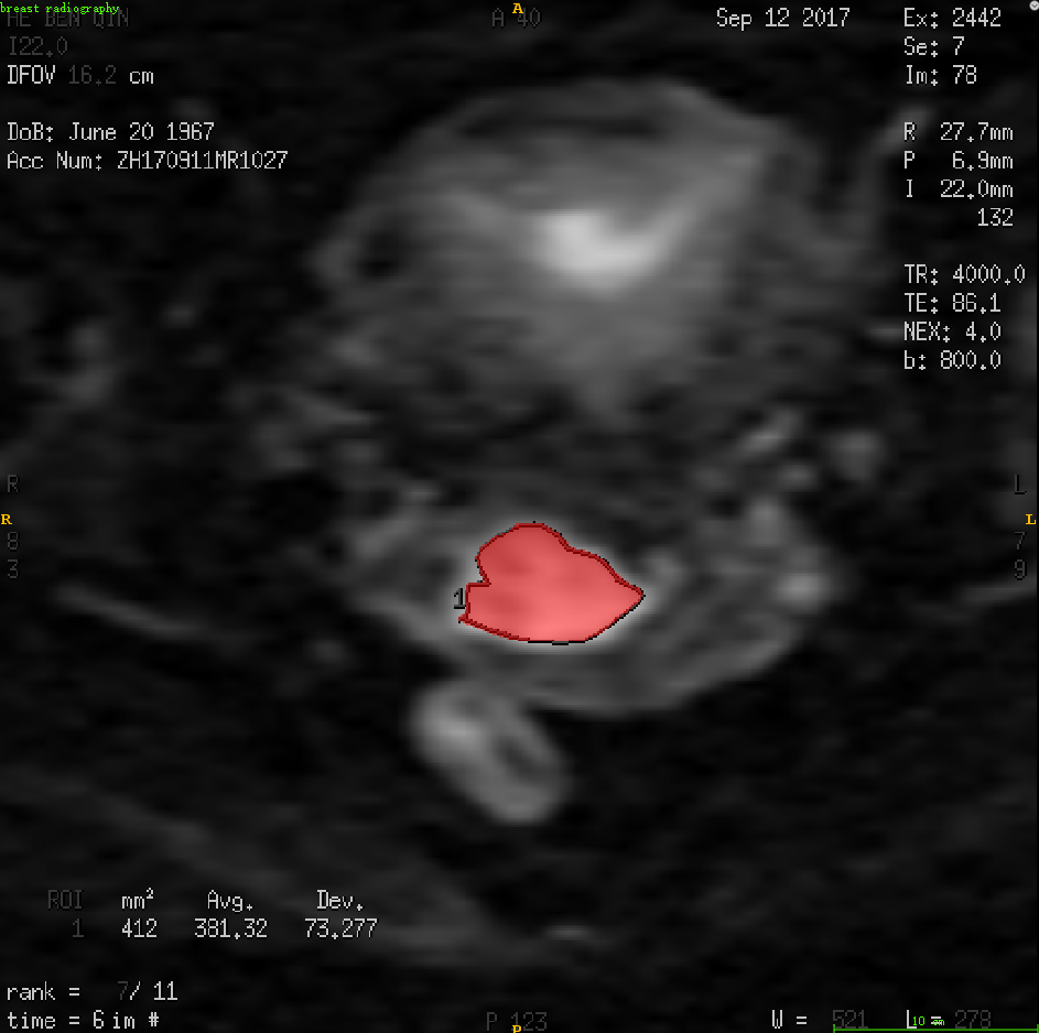
IVIM-DWI measurement, on Fifure# 1A (b = 800 s/mm2),
two radiologists drew ROI three times to get the values on the maps of D, D*
and f, respectively (Figure#1B-1D). Texture analysis, on Figure#1E, the
radiologists drew the ROI at the maximum area of the lesion in IVIM-DWI.