3680
Diffusion-weighted Imaging of Cervical Cancer Using Reduced-field-of-view, Readout-segmented, and Single-shot Echo-planar Imaging at 3T1West China Second University Hospital, Chengdu, China, 2West China Sencond University Hospital, Chengdu, China, 3MR Collaboration, Siemens Healthcare Ltd., Shanghai, China, 4MR Application Predevelopment, Siemens Healthcare GmbH, Erlangen, Germany
Synopsis
This study explored and compared the performance of reduced-FOV diffusion-weighted imaging (rFOV DWI), readout-segmented echo-planar-imaging (rs-EPI) DWI, and conventional single-shot (ss)-EPI DWI techniques for diagnosis of cervical cancer. Image quality was assessed by two radiologists; rFOV DWI and rs-EPI DWI achieved significantly better image quality and clinical utility at both b=50 s/mm2 and b=1000 s/mm2 compared with ss-EPI DWI, and rFOV DWI outperformed rs-EPI DWI in terms of artifacts, overall image quality, and clinical utility.
Introduction
Diffusion-weighted imaging (DWI) is an integral component of multi-parametric magnetic resonance imaging (MRI) of the cervical cancer. In high-field-strength (3T) imaging, the widely used ss-EPI technique is hampered by the presence of artifacts such as geometric distortion or susceptibility caused by B0 field variations and off-resonance effects. The rs-EPI DWI allows the use of shorter echo spacing in EPI echo sequences, thus reducing T2* blurring effects and obtaining higher resolution [1]. The rFOV DWI uses two-dimensional spatially selective excitation pulses to reduce the size of the FOV along the phase-encoding direction and thus shortens the length of the EPI echo sequence, thereby reducing geometric distortion and improving resolution [2]. However, a comparison between these three applications for diagnosis of cervical cancer has not been performed. The aim of this study was to determine the image quality and clinical utility of these three techniques applied to MRI of cervical cancer at 3T MRI.Materials and Methods
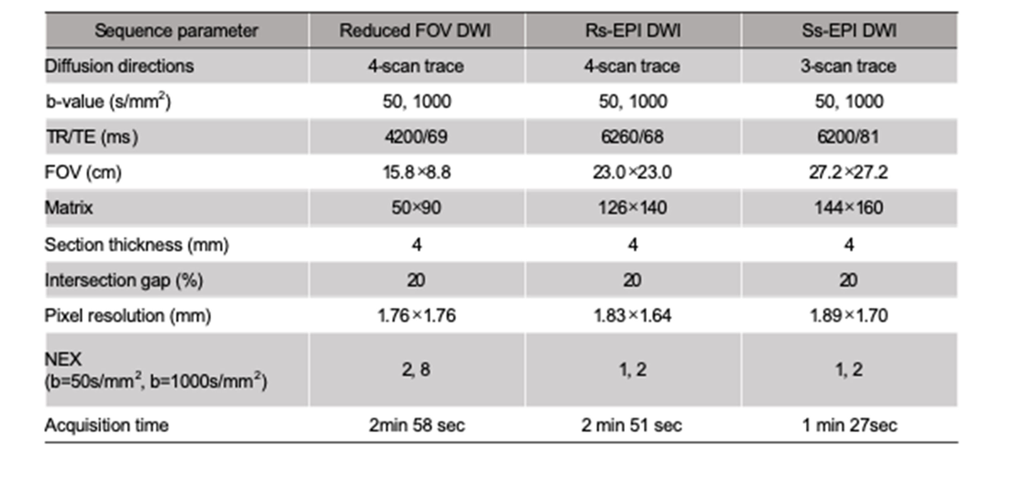
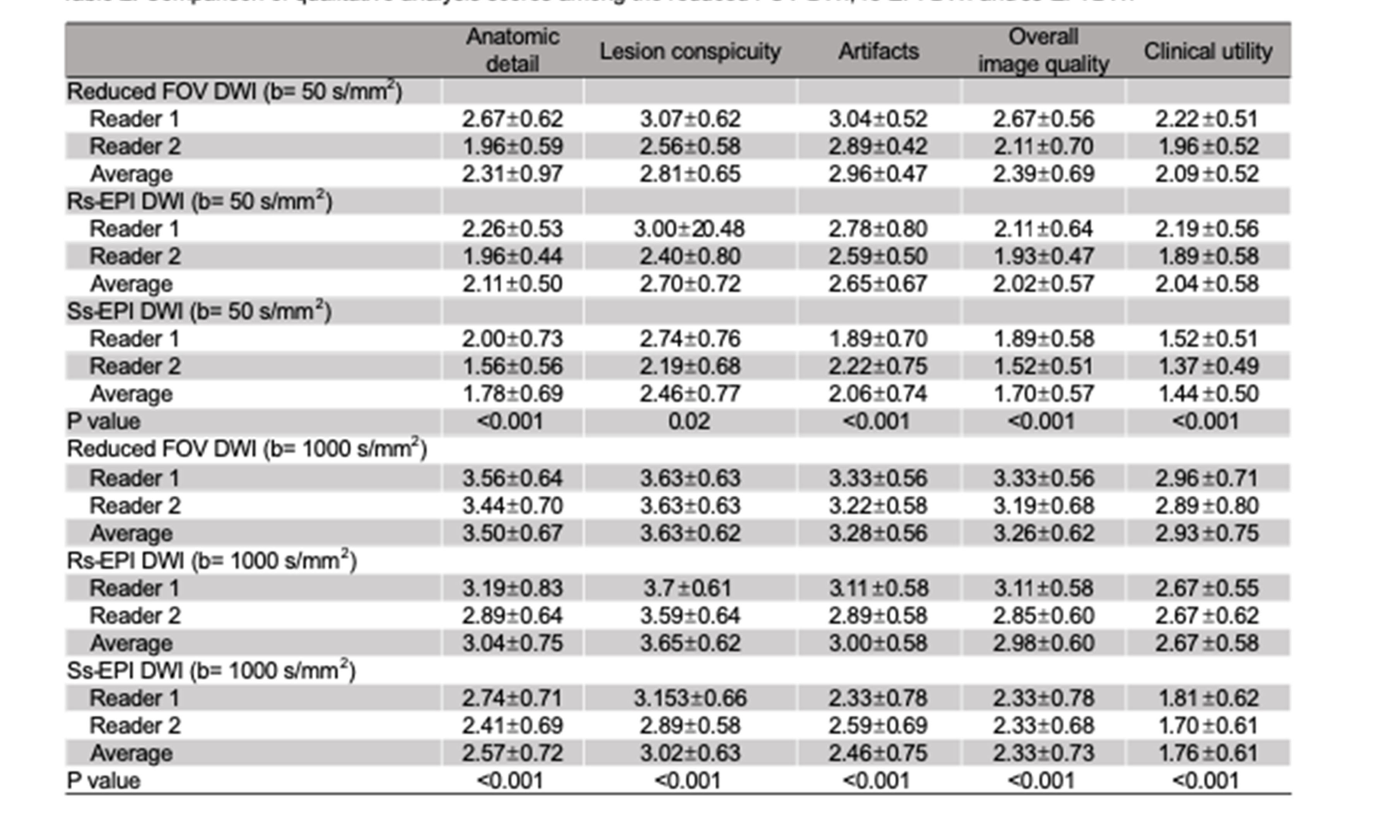
This study included 27 patients (mean age, 53.6 years) with biopsy-proven cervical cancer who underwent pelvic MRI before therapy on a 3T system (MAGNETOM Skyra, Siemens Healthcare, Erlangen, Germany), including rFOV DWI, rs-EPI DWI, and ss-EPI DWI. Acquisition parameters for each sequence are shown in Table 1. Two experienced radiologists independently assessed resulting images from each sequence on a 4-point scale (1 = poor, 2 = fair, 3 = good, 4 = excellent) for the following: anatomic detail, lesion conspicuity, artifact identification, overall image quality, and clinical utility. Clinical utility assessment (e.g. parametrial invasion) was determined by whether a given DWI method agreed with previous diagnosis made based on T1-weighted images, T2-weighted images, and dynamic enhanced images. Quantitative analyses were performed by manually placing a region of interest on the observed tumor and the apparent diffusion coefficient (ADC) was obtained. The statistical analysis included the Friedman test and Mann-Whitney test for comparisons of image quality and ADC values.Results
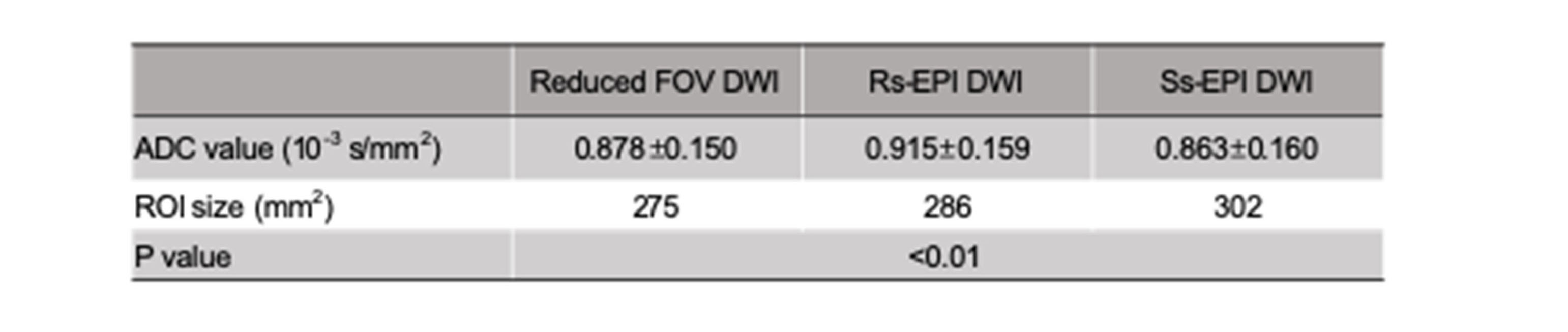
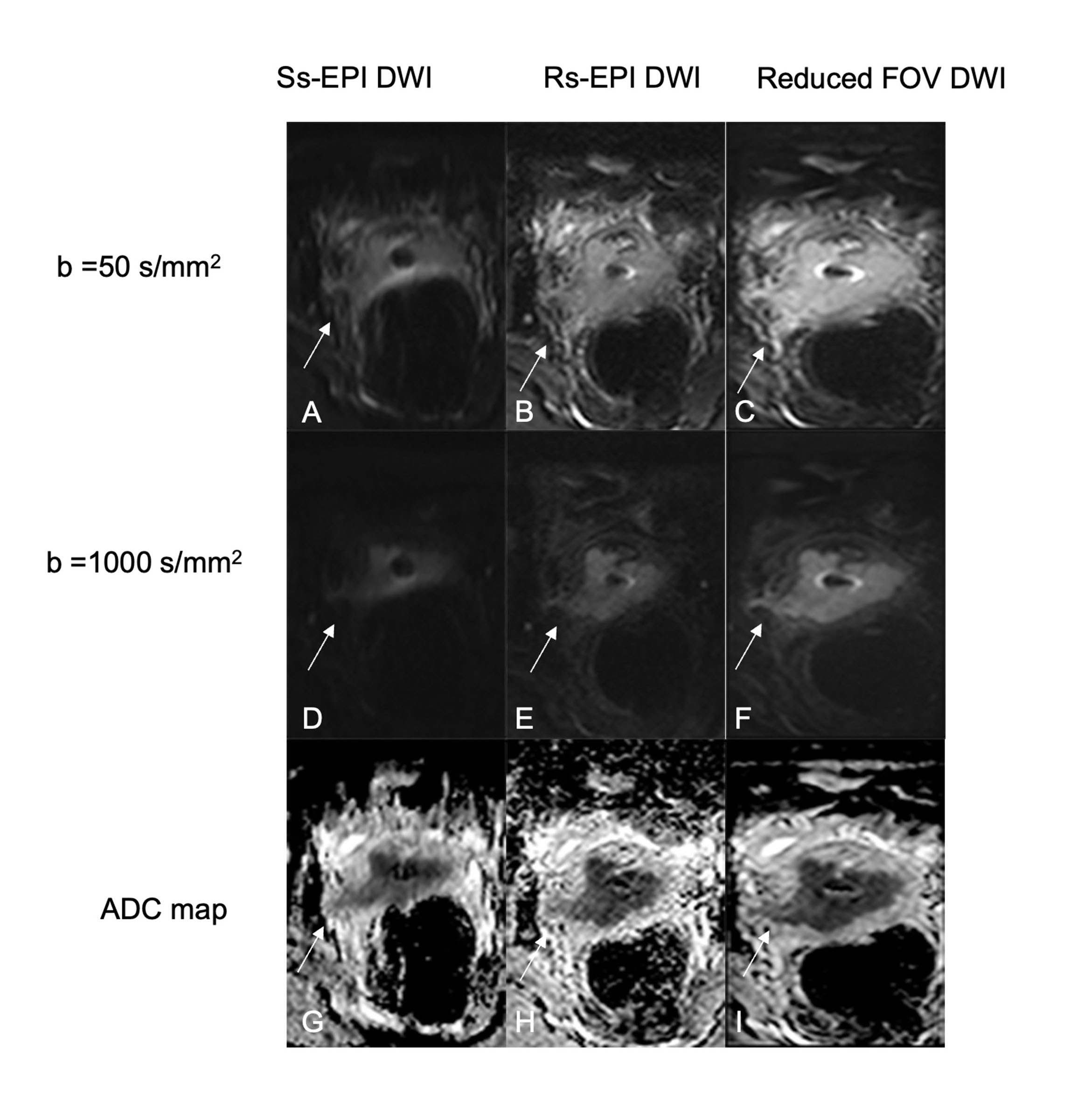
Example image results for all three DWI sequences (at b=50 s/mm2 and b=1000 s/mm2) are shown in Figure 1. Table 2 presents summary results over scoring for all three DWI sequences and both readers. For both observers, rFOV DWI and rs-EPI DWI at both b=50 s/mm2 and b=1000 s/mm2 had significantly higher scores for anatomic detail, lesion conspicuity, artifact identification, overall image quality, and clinical utility compared to ss-EPI DWI (P<0.05). Significantly lower number of artifacts, better anatomic detail, higher overall image quality, and better clinical utility were observed in rFOV DWI compared with rs-EPI DWI (P<0.05). For the anatomic detail score at b=50 s/mm2, reader 2 found high agreement between rFOV DWI and rs-EPI DWI (P<0.001). The result of reader 1 for the lesion conspicuity score at b=1000 s/mm2 was slightly higher for rs-EPI DWI than for rFOV DWI. Interobserver agreement was fair to excellent among the three DWI sequences (κ = 0.21–0.63 at b = 0 s/mm2 and κ = 0.28–0.85 at b = 1000 s/mm2, Table 3). There was a significant difference in the mean tumor ADC values among the three sequences (P<0.01) (Table 4).Discussion
In this study, we compared rFOV DWI, rs-EPI DWI, and ss-EPI DWI in order to evaluate the image quality and parametrial invasion in endometrial cancer. DWI in the uterus suffers from high susceptibility and motion artifacts. In agreement with previous research findings [3-5], our results indicated that rFOV DWI and rs-EPI DWI are both preferred to ss-EPI DWI due to their better anatomic detail, lesion conspicuity, artifact identification, overall image quality, and clinical utility. This was a result of side-by-side comparison of the three DWI sequences in which both readers quantitatively rated rFOV DWI and rs-EPI DWI higher than ss-EPI DWI. Although one reader's results show that the anatomic detail of the two sequences is very close at b=50 s/mm2, the combined results of the two readers still indicate that the anatomic detail of rFOV DWI is better than rs-EPI DWI. Lesion conspicuity was slightly lower for rFOV DWI than for rs-EPI DWI, which may be a result of interpolation was used for rs-EPI, but not for rFOV.Conclusion
Reduced-FOV DWI and rs-EPI DWI showed improved image quality and clinical utility compared with conventional DWI. Reduced-FOV DWI also outperformed rs-EPI DWI in terms of image quality and clinical utility.Acknowledgements
No acknowledgement found.References
[1] Porter DA, Heidemann RM. High resolution diffusion-weighted imaging using readout-segmented echo-planar imaging, parallel imaging and a two-dimensional navigator-based reacquisition. Magn Reson Med. 2009;62:468–475.
[2] Thierfelder KM, Scherr MK, Notohamiprodjo M, et al. Diffusion-weighted MRI of the prostate: advantages of Zoomed EPI with parallel-transmit-accelerated 2D-selective excitation imaging. Eur Radiol. 2014;24:3233–3241.
[3] Hwang J, Hong SS, Kim HJ, et al. Reduced field-of-view diffusive -weighted MRI in patients with cervical cancer. Br J Radiol. 2018; 20170864.
[4] Takeuchi M, Matsuzaki K, Bando Y et al. educed field-of-view diffusion-weighted MR imaging for assessing the local extent of uterine cervical cancer. Acta Radiol. 2020;61:267-275.
[5] Yeom KW, Holdsworth SJ, Van AT, et al. Comparison of read-out segmented echo-planar imaging (EPI) and single-shot in clinical application of diffusion-weighted imaging of the pediatric brain. AJR Am J Roentgenol. 2013; 200: 437–443.
Figures