3666
Preliminary Study on Monitoring Drug Resistance of Colon Cancer with Intravoxel Incoherent Motion MRI In Vivo1Medical Imaging Department, Nansha Hospital, Guangzhou First People's Hospital, School of Medicine, South China University of Technology, Guangzhou, China, 2Ultrasound Imaging Department, Longgang District People’s Hospital, Shenzhen, China, 3Department of Radiology, The Second Affiliated Hospital of Guangzhou University of Chinese Medicine, Guangdong Provincial Hospital of Traditional Chinese Medicine, Guangzhou, China, 4Department of Pathology, Cancer Center, Sun Yat-sen University, Guangzhou, China, 5Department of Pathology, Nansha Hospital, Guangzhou First People's Hospital, School of Medicine, South China University of Technology, Guangzhou, China, 6Medical Record Department, Nansha Hospital, Guangzhou First People's Hospital, School of Medicine, South China University of Technology, Guangzhou, China, 7MR Scientific Marketing, SIEMENS Healthcare Ltd., Guangzhou, China
Synopsis
This study has demonstrated that ADC value of DWI with single-exponential model and diffusion coefficient D of IVIM are valuable for discriminating 5-FU-responsive and 5-FU-resistant colon cancer in vivo. IVIM is expected to be a simple and practical way to quantitatively monitor tumor resistance in vivo, and ADC &D value may be an imaging biomarker.
Purpose
To investigate the role of intravoxel incoherent motion-diffusion weighted imaging (IVIM-DWI) in evaluating the drug resistance in colon cancer xenografts and to explore the possible biomarkers.Methods
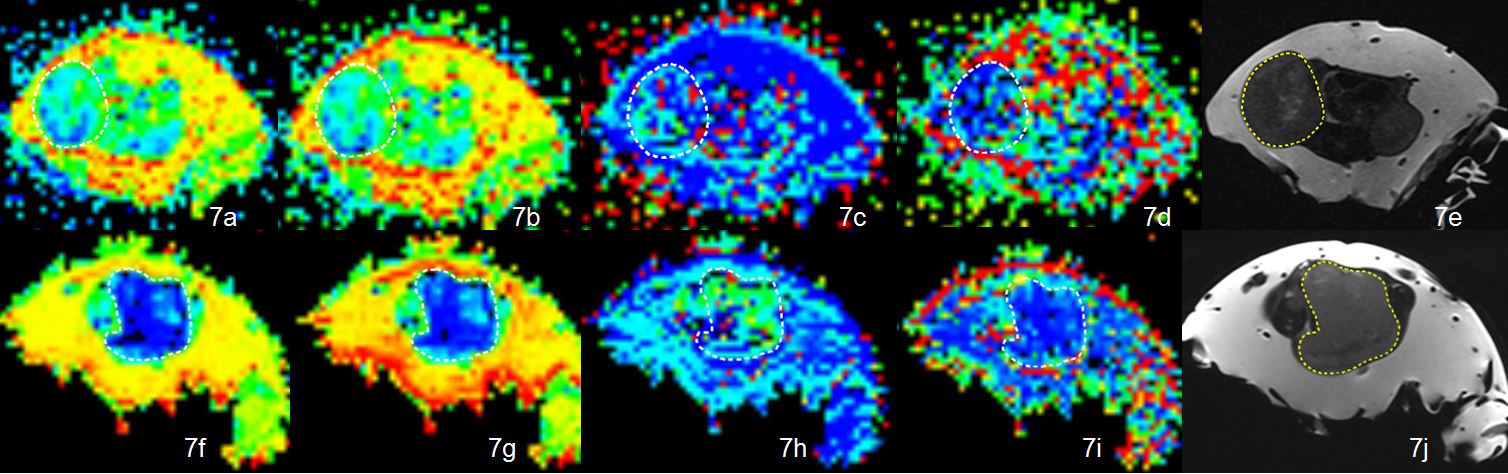
10 healthy BALB/c nude mice were divided into 2 groups randomly(5 each),and they have been injected SW480 or SW480/5-FU cell suspensions respectively.MR imaging was performed on a 3T MR scanner (MAGNETOM Skyra; Siemens Healthcare, Erlangen, Germany) with an 8-channel mouse coil (Chenguang Medical Technology Co. Shanghai, China). T2-weighted fast spin-echo(TSE) images and IVIM sequences were obtained for all the mice. The main scan parameters of MR sequences are as following: T2WI (TSE): TR/TE=4500ms/110ms, slice thickness=2mm, FOV=128mm ×128mm, reconstruction matrix = 512 × 512, acquisition time = 2min 2s, axial, coronal, sagittal imaging (Fig 2).DWI: Axial imaging was acquired with single-shot echo-planar imaging(EPI), isotropic diffusion-sensitive gradient field and 8 b-values (0, 50, 100, 150, 200, 400, 800, 1200 s/mm2), TR=3400ms, TE=60ms, FOV=220mm × 220mm, NEX=3, slice thickness=2mm, acquisition matrix=220×220, acquisition time=13min 39s . Multi-b value DWI data were analyzed with MITK Diffusion (Version 2013.03.00). The regions of interest (ROIs) were manually drawn in the largest section of the tumor, avoiding bleeding, cystic and necrotic areas by at least two radiologists independently with unknown tumor-related information. Each tumor was measured three times respectively by each radiologist. The average value measured by two radiologists was included in the analysis. The acquisition parameters include the ADC, D, D*, and f values of the tumor. The mean ROI size was 51.1 mm2 (range, 7.1–94.91 mm2) by observer 1 and 52.3 mm2 (range, 8.4–98.2mm2 ) by observer 2.
Then their tumors were removed to detect the protein expression of P-gp, MPR1, PKC, cell morphology and necrosis, apoptosis, and drug resistance.
Results
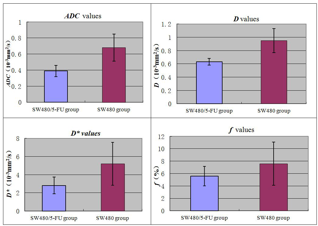
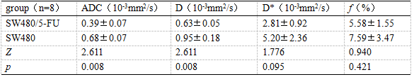
The ADC values and the values of D were significantly higher in SW480 group and there was no statistical difference of D*, f value between the two groups. The cell nucleus was larger and the cells arranged more closely in SW480/5-FU group. The necrosis degree and apoptosis rate of the tumor had no significant difference between the two groups. The protein expressions of P-gp, MPR1, PKC were significantly higher in SW480/5-Fu group (P<0.05).Conclusion
The ADC value of DWI and diffusion coefficient D of IVIM were moderately valuable for discriminating 5-FU-response and resistance of colon cancer in vivo. IVIM is expected to be a convenient and practical way to quantitatively monitor tumor resistance in vivo, and ADC & D value would be effective imaging biomarkers for tumor analysis.Discussion
Our work has revealed a mechanism of lower D values with IVIM in 5-FU-resistant colon cancer tissue. The colon cancer cells of 5-FU-resistant have a high cell turnover with more cell division resulting in densely packed cells and enlargement nucleus so that prevent water molecules from diffusing. It is expected to quantify tumour hypercellularity in vivo.For more perfusion weighted parameters f & D*, f represents the contribution of water moving in capillaries, and D* represents the diffusion within the microcirculation. In theory,the IVIM perfusion method offers a way to study microcirculatory blood-flow properties and to monitor early response of cancer with antiangiogenic treatment or vascular targeting agents treatment in vivo. Anti-angiogenic drugs control the growth of tumors by affecting the vascular endothelial growth factor family (VEGF) of pathway. Cui et al. found that f and D* were significantly reduced compared with the control group 1 day after anti-angiogenesis treatment in a mouse model of nasopharyngeal carcinoma. Vascular targeting agents are designed to cause rapid selective closure of tumor blood vessels. Joo et al. found that f, D* and fD* were significantly reduced 4 hours after administration of a vascular disrupting agent to a rabbit model of liver tumor. Subsequent observations showed that the decrease in f and fD* at 4 h was inversely related to the increase in tumor size on day 7 after treatment.In this study, the f-value and D* value of the response colon cancer group(SW480) were higher than those of the 5-FU-resistant colon cancer group(SW480/5-FU), but there was no significant difference between the two groups (P >0.05). Tumor tissue sections showed there was no significant difference of intratumoral necrosis between 5-FU-responsive and resistant groups. These results suggest that the perfusion of the 5-FU-resistant cancer tissue is reduced, but not to the extent that it causes ischemic necrosis of the tumor. 5-FU-resistant colon cancer cells have improved viability in a low perfusion state.Acknowledgements
No acknowledgement found.References
1. Murtz P, Sprinkart AM, Reick M, et al. Accurate IVIM model-based liver lesion characterisation can be achieved with only three b-value DWI. Eur Radiol. 2018;28(10):4418-4428.
2. Xu Y, Xu Q, Sun H, et al. Could IVIM and ADC help in predicting the KRAS status in patients with rectal cancer? Eur Radiol. 2018;28(7):3059-3065.
3. Mannelli L, Nougaret S, Vargas HA, et al. Advances in diffusion-weighted imaging. Radiol Clin North Am. 2015;53(3):569-81.
4. Jerome NP, Boult JKR, Orton MR, et al. Characterisation of fibrosis in chemically-induced rat mammary carcinomas using multi-modal endogenous contrast MRI on a 1.5T clinical platform. Eur Radiol. 2018;28(4):1642-1653.
5. De Robertis R, Tinazzi Martini P, Demozzi E, et al. Diffusion-weighted imaging of pancreatic cancer. World J Radiol. 2015;7(10):319-28.
6. Iima M, Kataoka M, Kanao S, et al. Intravoxel Incoherent Motion and Quantitative Non-Gaussian Diffusion MR Imaging: Evaluation of the Diagnostic and Prognostic Value of Several Markers of Malignant and Benign Breast Lesions. Radiology. 2018;287(2):432-441.
7. Cho GY, Moy L, Kim SG, et al. Evaluation of breast cancer using intravoxel incoherent motion (IVIM) histogram analysis: comparison with malignant status, histological subtype, and molecular prognostic factors. Eur Radiol. 2016;26(8):2547-58.
8. Rong D, Mao Y, Hu W, et al. Intravoxel incoherent motion magnetic resonance imaging for differentiating metastatic and non-metastatic lymph nodes in pancreatic ductal adenocarcinoma. Eur Radiol. 2018;28(7):2781-2789.
Figures
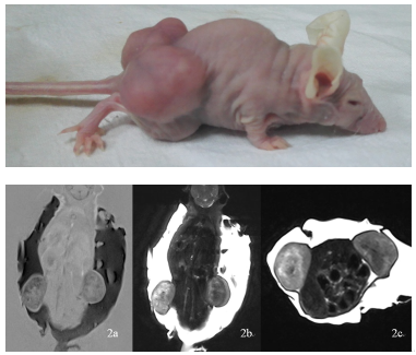
Nude mice xenograft of SW480.
MR imaging T1WI coronal(2a),T2WI coronal(2b) and T2WI axial slices(2c).