3654
Reducing Rician noise bias in axial-symmetric Diffusion Kurtosis Imaging and biophysical tissue models1Institute of Systems Neuroscience, University Medical Center Hamburg-Eppendorf, Hamburg, Germany, 2Weierstrass Institute for Applied Analysis and Stochastics, Berlin, Germany, 3Department of Neurophysics, Max Planck Institute for Human Cognitive and Brain Sciences, Leipzig, Germany
Synopsis
Five out of eight axial-symmetric Diffusion Kurtosis Imaging (AxDKI) parameters are directly related to biophysical microstructure parameters including intra- and extra-axonal diffusivities, fiber dispersion and axonal-water fraction. Their estimation, however, is biased at small signal-to-noise ratios (SNR). Here, based on simulations, we investigated the Rician noise bias’s effect and its correction (RBC) on estimated AxDKI and biophysical parameters at varying SNRs for the standard and AxDKI model. Our study suggests AxDKI to be better than standard DKI, here least biased AxDKI estimators were produced with RBC while for biophysical parameters results were branch-dependent (SNR≥23 with RBC and SNR≥33 without RBC).
Introduction:
Diffusion Kurtosis Imaging (DKI) has recently attracted additional attention in form of the axial-symmetric DKI (AxDKI) signal model1 and its relation to the biophysical parameters intra- and extra-axonal diffusivity, fiber dispersion and axonal water fraction2,3. The axial-symmetric DKI model assumes axial-symmetrically distributed axons around an axis of symmetry and reduces the number of model parameters from 22 for standard DKI to 8, making it more robust and less data demanding. DKI model parameters are typically estimated from MRI magnitude images which are contaminated with Rician noise4. While the influence of the resulting Rician bias and its correction on parameter estimates was investigated within the standard DKI model5–8, it is currently unknown how Rician noise bias correction (RBC) can help to improve parameter estimation in axial-symmetric DKI and the associated biophysical parameters. Here, the efficacy of a newly implemented RBC algorithm (using quasi-likelihood estimators9) on axial-symmetric DKI parameter estimation and the consequent computation of the biophysical parameters was investigated based on a simulation study with in-vivo white matter datasets.Methods:
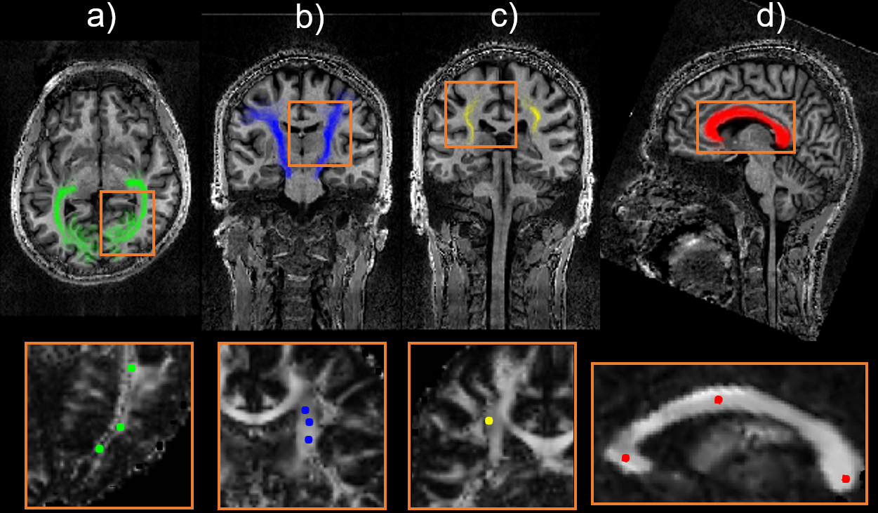
Twelve noise-free diffusion datasets were simulated from parameters of the standard DKI model, estimated from a diffusion dataset from a healthy volunteer11 within selected ROIs, see Figure 1. Data were simulated using a three-shell diffusion sequence11. Corresponding noisy data were obtained for varying SNR=1 to 100; 2500 samples per SNR were drawn from a complex Gaussian distribution $$$N(0,σ)$$$ with $$$\sigma = \sqrt2 \frac{S_0}{SNR} $$$. From the magnitude data, all parameters from the AxDKI and the standard DKI model12 were estimated and biophysically-relevant parameters $$$θ=\{D_\parallel,D_\perp,W_\parallel,W_\perp,\overline{W}\}$$$ were compared to the noise-free ground truth (GT) by computing the bias $$$100 \cdot \frac{\vert GT-Fit Results\vert }{GT}$$$. DKI parameter estimation was done in a least squares (no RBC) or quasi-likelihood (RBC) approach as in9 using $$$S(b,\vec{g_i})$$$ or the expectation value $$$μ(S(b,\vec{g_i}), σ)$$$ of the Rician distributed data, respectively. Here $$$S(b,\vec{g_i})$$$ is the signal model (diffusion weighting b and i'th diffusion gradient $$$\vec{g_i}$$$). From $$$θ$$$ the axon dispersion $$$κ $$$ was determined solving the optimization problem detailed in3,13. From $$$θ$$$ and $$$κ$$$ the biophysical parameters f (axonal water fraction), $$$D_a$$$ (parallel intra-axonal diffusivity), $$$D_{e,\perp}$$$ and $$$D_{e,\parallel}$$$ (parallel and perpendicular extra-axonal diffusivity) can be calculated using analytical expressions3,13. The problem is generally degenerated such that there is more than one set of solutions (“branches”). The implementations are Matlab-based and freely available online within the ACID Toolbox (http://www.diffusiontools.com/).Results:
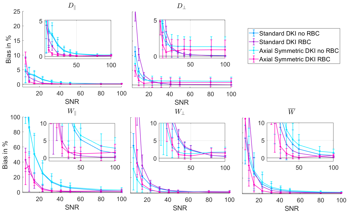
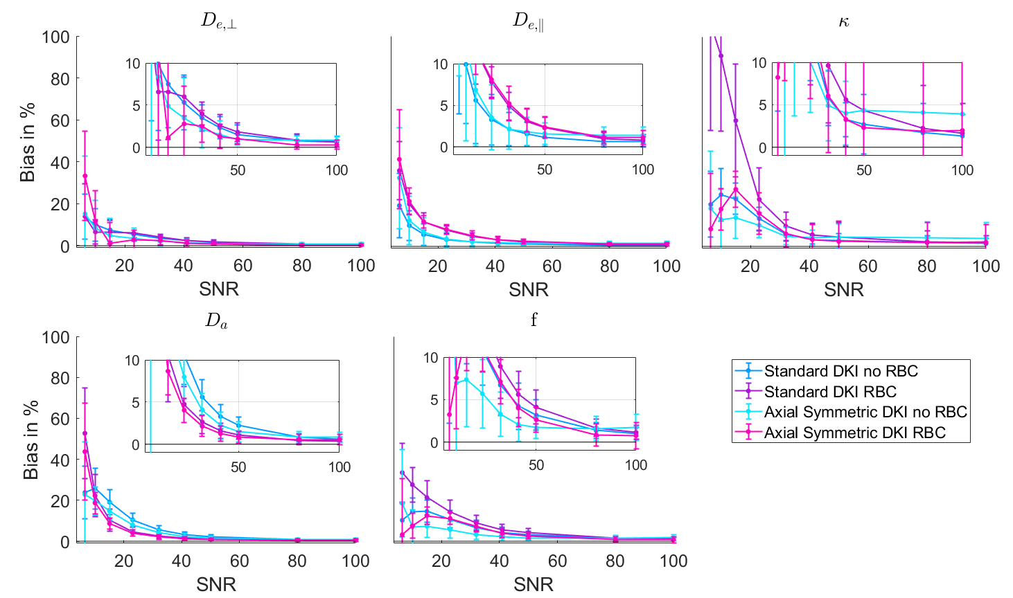
θ parameters: The diffusion parameters $$$D_{\perp}$$$ and $$$D_{\parallel}$$$ were generally less biased compared to the kurtosis parameters (y axes, Figure 2). RBC reduced the bias for both signal models for the parallel parameters ($$$D_{\parallel}$$$ and $$$W_{\parallel}$$$). The estimation bias for the perpendicular parameters $$$D_{\perp}$$$ and $$$W_{\perp}$$$ was lower in the AxDKI model compared to the standard model with a residual bias ≈1% for high SNRs. For $$$\overline{W}$$$ combining AxDKI with RBC produced the lowest bias. Surprisingly, for low SNRs the RBC increased the bias, except for $$$W_{\parallel}$$$ and the axial-symmetric fit for $$$D_{\perp}$$$ (Figure 2).Biophysical parameters branch one: RBC reduced the bias for $$$D_a$$$ regardless of the used model (Figure 3). $$$D_{e,\parallel}$$$, $$$D_{e,\perp}$$$, f and $$$κ $$$ were best estimated by AxDKI, surprisingly, RBC increased the bias in this case.
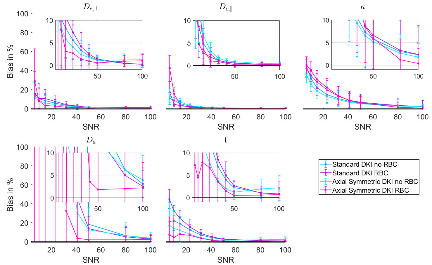
Biophysical parameters branch two: Estimation of $$$D_a$$$ proved to be unstable for low SNRs showing high biases due to outliers (Figure 4). For the other parameters, the axial-symmetric DKI fit with RBC produced the overall least biased estimators for high SNRs≥50 whilst showing the strongest bias for the SNR=6.5 datapoint for $$$D_{e,\parallel}$$$ and $$$D_{e,\perp}$$$. For SNRs up to 50, $$$κ$$$ is best estimated with the AxDKI model without RBC.
Discussion:
The RBC reduced the AxDKI parameter estimation bias predominantly in the parallel parameters $$$D_{\parallel}$$$, $$$W_{\parallel}$$$ and $$$\overline{W}$$$, less for the perpendicular parameters $$$D_{\perp}$$$ and $$$W_{\perp}$$$. For the biophysical parameters the RBC showed branch-dependent performance, improving only $$$D_a$$$ (intra-axonal diffusivity parallel to axons) for branch one and all parameters for branch two. Overall, the axial-symmetric model produced the least biased AxDKI estimators when used with RBC, providing a bias <5% at an SNR≥23, a bias <5% at an SNR≥33 when used without RBC for the biophysical parameters of branch one and a bias <5% at an SNR≥50 for the biophysical parameters of branch two.The observed pattern can be understood in terms of the underlying tissue structure. Measurements parallel to the main fiber direction are most compromised by Rician noise because signal attenuation is strongest making RBC at lower SNR more important. Accordingly, the parallel parameters were most efficiently estimated by the RBC fits. The superiority of the axial-symmetric model for the perpendicular DKI parameters and $$$\overline{W}$$$ could be caused by a more robust parameter estimation in case of these averaged variables. The higher SNR requirement to reduce the overall estimation bias for the biophysical parameters of branch two is driven by the outliers for $$$D_a$$$ for very low SNRs and an overall higher bias for $$$κ$$$ .
Conclusion
Our results suggest that axial-symmetric DKI is well suited to estimate kurtosis and biophysical parameters, although it is a reduced model. RBC works best for the parallel parameters. A possible limitation to axial-symmetric DKI is the violation of the symmetry assumption, possibly in gray brain matter.Acknowledgements
This work was supported by the German Research Foundation (DFG Priority Program 2041 "Computational Connectomics”, [AL 1156/2-1;GE 2967/1-1; MO 2397/5-1; MO 2249/3–1], by the Emmy Noether Stipend: MO 2397/4-1) and by the BMBF (01EW1711A and B) in the framework of ERA-NET NEURON.References
1. Hansen, B., Shemesh, N. & Jespersen, S. N. Fast imaging of mean, axial and radial diffusion kurtosis. NeuroImage 142, 381–393 (2016).
2. Fieremans, E., Jensen, J. H. & Helpern, J. A. White matter characterization with diffusional kurtosis imaging. NeuroImage 58, 177–188 (2011).
3. Jespersen, S. N., Olesen, J. L., Hansen, B. & Shemesh, N. Diffusion time dependence of microstructural parameters in fixed spinal cord. NeuroImage 182, 329–342 (2018).
4. Gudbjartsson, H. & Patz, S. The rician distribution of noisy mri data. Magn. Reson. Med. 34, 910–914 (1995).
5. Veraart, J., Hecke, W. V. & Sijbers, J. Constrained maximum likelihood estimation of the diffusion kurtosis tensor using a Rician noise model. Magn. Reson. Med. 66, 678–686 (2011).
6. Veraart, J. et al. Comprehensive framework for accurate diffusion MRI parameter estimation. Magn. Reson. Med. 70, 972–984 (2013).
7. Koay, C. G., Özarslan, E. & Basser, P. J. A signal transformational framework for breaking the noise floor and its applications in MRI. J. Magn. Reson. 197, 108–119 (2009).
8. André, E. D. et al. Influence of Noise Correction on Intra- and Inter-Subject Variability of Quantitative Metrics in Diffusion Kurtosis Imaging. PLoS ONE 9, (2014).
9. Polzehl, J. & Tabelow, K. Low SNR in Diffusion MRI Models. J. Am. Stat. Assoc. 111, 1480–1490 (2016).
10. Eickhoff, S. B. et al. A new SPM toolbox for combining probabilistic cytoarchitectonic maps and functional imaging data. NeuroImage 25, 1325–1335 (2005).
11. Mohammadi, S. & Callaghan, M. F. Towards in vivo g-ratio mapping using MRI: Unifying myelin and diffusion imaging. J. Neurosci. Methods 108990 (2020) doi:10.1016/j.jneumeth.2020.108990.
12. Tabesh, A., Jensen, J. H., Ardekani, B. A. & Helpern, J. A. Estimation of tensors and tensor-derived measures in diffusional kurtosis imaging. Magn. Reson. Med. 65, 823–836 (2011).
13. Novikov, D. S., Veraart, J., Jelescu, I. O. & Fieremans, E. Rotationally-invariant mapping of scalar and orientational metrics of neuronal microstructure with diffusion MRI. NeuroImage 174, 518–538 (2018).
Figures