3633
Early Cardiac Involvement Detected by CMR Feature Tracking in Idiopathic Inflammatory Myopathy with Preserved Ejection Fraction1the first affiliated hospital of Nanjing medical university, Nanjing, China
Synopsis
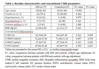
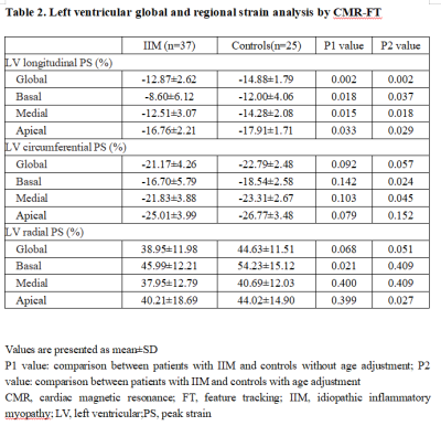
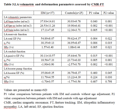
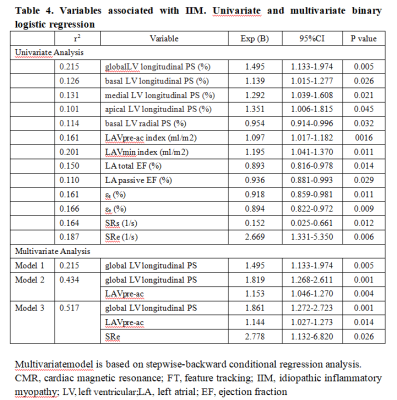
We applied CMR-FT technique to evaluate LV and LA performance in IIM patients. The main findings were as follow: (1) compared with controls, the damaged LV strain in IIM patients mainly involved global and regional LV PS in longitudinal direction; (2) LA reservoir function and conduit function were impaired in IIM patients; (3) LA volumetric parameters and function parameters showed significant difference between IIM patients and the controls. (4) Combination of global LV longitudinal PS, LAVpre-ac index and Apical LV circumferential PS can improve diagnostic accuracy. Therefore, CMR-FT can help early diagnosis of cardiac involvement of IIM patients.
Introduction
Cardiac involvement is common in idiopathic inflammatory myopathy (IIM) but often subclinical1. At present, myocardial injury of IIM could not be detected sensitively or specifically. Thus, a valuable technique is needed. The aim of this study was to assess cardiac involvement in IIM patients by cardiac magnetic resonance feature tracking (CMR-FT).Methods
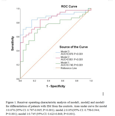
Thirty-seven IIM patients and 25 controls were studied at 3.0 Tesla. All participants underwent cine CMR imaging on a 3.0T whole-body scanner (MAGNETOM Skyra, Siemens Healthcare, Erlangen, Germany) with an 18-channel phase-array coil using ECG gating. The b-SSFP sequences with breath-hold were performed to obtain cine CMR images, comprising a stack of contiguous parallel short-axis slices covering the entire LV from base to apex. The typical parameters were as follows: field of view (FOV), 340-380 mm; repetition time (TR) /echo time (TE), 3.4/1.4 ms; matrix size, 208×188 mm; voxel size, 1.6×1.6×8.0 mm; bandwidth, 962 Hz/Px; flip angle (FA), 47°; slice thickness, 8 mm; inter slice gap, 2 mm. Three LV long-axis slice (2-, 3-, and 4-chamber views) images were also obtained. The left ventricular functional parameters such as volume and EF were measured. Global and regional (basal, medial, apical) left ventricle (LV) peak strain (PS) in radial, circumferential and longitudinal directions were derived from balanced steady state free precession (b-SSFP) cine images. Left atrial (LA) volumes were assessed at three time-points of the cardiac cycle. LA longitudinal strain and strain rate (SR) parameters were analyzed using 4- and 2-chamber views including LA reservoir function (total ejection fraction [EF], total strain [εs], peak positive SR [SRs]), LA conduit function (passive EF, passive strain [εe], peak early negative SR [SRe]) and LA booster pump function (booster EF, active strain [εa], late peak negative SR [SRa]) were assessed, respectively. Three models were assessed: model 1, based on global LV longitudinal PS; model 2, based on global LV longitudinal PS and LAVpre-ac indexed; model 3, based on global LV longitudinal PS, LAVpre-ac indexed and SRe. Unpaired Student’s t test, Chi square test or fisher exact-test, logistic regression, receiver operating characteristic curve and intra-class correlation coefficient analysis were applied for statistical analyses.Results
IIM patients showed significantly reduced global and regional LVPS in longitudinal direction (all P<0.05)(Table 2). Compared with controls, LA reservoir (LA total EF, εs, SRs) and conduit function (LA passive EF, εe, SRe) were significantly impaired in IIM patients (all p < 0.05) (Table 3). All models were useful for diagnosis, and model 3 showed the highest diagnostic accuracy (AUC, 0.876; sensitivity, 83.8%; specificity, 84%)(Figure 1). The global LV longitudinal PS was an independent predictor of IIM. By Pearson’s correlation analysis, the LV global radial, circumferential and longitudinal PS were all correlated to LVEF in IIM patients (R=0.526, P<0.001 vs. R=-0.514, P<0.001 vs. R=-0.288, P=0.023). Observer variability was excellent for all strain and SR parameters on an intra- and inter-observer level as determined by intraclass correlation coefficient analyses.Discussion
Compared with the controls, the magnitude of global and regional LV longitudinal PS were decreased in IIM patients with preserved LVEF, which is consistent with previous results 2,3. This finding may explain the impairment of LV deformation detected by CMR-FT prior to conventional LVEF in IIM patients. LV longitudinal PS represents the contraction of the longitudinal myocardial fibers in the LV subendocardium, which is the most susceptible layer to various stressors, longitudinal strain parameters therefore have the sensitivity to detect subtle systolic dysfunction before conventional changes in LVEF 4. Previous studies have demonstrated that IIM patients showed impaired LV myocardial microvascular dysfunction, and that the abnormal LV myocardial deformation was associated with microvascular dysfunction in other diseases2,5. Thus, we presume that the magnitude reduction in longitudinal myocardial strain of IIM patients in this study may be related to LV myocardial microvascular ischemia.The frequency of LV diastolic dysfunction in IIM patients was 42% 1. LA reservoir and conduit function reflect left ventricular diastolic dysfunction, while impaired booster pump function reflect dysfunction of left atrial contraction 6. The reservoir and conduit functions make the main contribution to LV filling during early diastole, while the booster pump function is the basis for active LV filling during late diastole 7. In this study, LA reservoir function and LA conduit function showed significant difference between controls and IIM patients. The suggested main mechanisms of cardiovascular involvement in IIM include the effects of traditional cardiovascular risk factors, systemic and local inflammation of the disease. Hypertension, diabetes mellitus and ischemia heart disease are more prevalent in IIM patients than in general population8. Increased cardiac load forces adaptive myocardial remodelling to uphold the perfusion of the peripheral tissues in IIM patients. Cardiac remodelling leads to hypertrophy and enlargement of heart chambers 9. Hence, in IIM patients, chronic pressure overload, LV hypertrophy, myocardial fibrosis could lead to elevated filling pressure, LV diastolic dysfunction, aggravated LV stiffness and LA remodelling.Conclusion
CMR-FT based LV and LA performance analysis reliably quantifies strain parameters, which could detect early cardiac involvement in IIM patients.Acknowledgements
noneReferences
1. Danko K, Ponyi A, Constantin T. Long-term survival of patients with idiopathic inflammatory myopathies according to clinical features. Medicine (Baltimore). 2004;83:35–42.
2. Wang J, Shi K, Xu HY, et al. Left Ventricular Deformation in Patients with Connective Tissue Disease: Evaluated by 3.0T Cardiac Magnetic Resonance Tissue Tracking. Sci Rep. 2019 Nov 29;9(1):17913.
3. HuberA T, Bravetti M, Lamy J, et al. Non-invasive differentiation of idiopathic inflammatory myopathy with cardiac involvement from acute viral myocarditis using cardiovascular magnetic resonance imaging T1 and T2 mapping. Journal of Cardiovascular Magnetic Resonance, 2018, 20(1):11.
4. Zhong Y, Bai W, Xie Q, Sun J, Tang H, Rao L. Cardiac function in patients with polymyositis or dermatomyositis: a three-dimensional speckle-tracking echocardiography study. Int J Cardiovasc Imaging. 2018 May;34(5):683-693. doi: 10.1007/s10554-017-1278-9. Epub 2017 Nov 22.
5. Jayakumar D, Zhang R, Wasserman A, et al. Cardiac Manifestations in Idiopathic Infammatory Myopathies: An Overview. Cardiology in review,2019, 27, 131–137.
6. Hoit BD. Left atrial size and function: role in prognosis. journal of the american college of cardiology, 2014, 63(6):493-505.
7. Li L , Chen X , Yin G , et al. Early detection of left atrial dysfunction assessed by CMR feature tracking in hypertensive patients. European Radiology, 2019.
8. Limaye VS, Lester S, Blumbergs P, Roberts-Thomson PJ. Idiopathic inflammatory myositis is associated with a high incidence of hypertension and diabetes mellitus. Int J Rheum Dis. 2010 May;13(2):132-7. doi: 10.1111/j.1756-185X.2010.01470.x.
9. Opinc AH, Makowski MA, Łukasik ZM, Makowska JS. Cardiovascular complications in patients with idiopathic inflammatory myopathies: does heart matter in idiopathic inflammatory myopathies? Heart Fail Rev. 2019 Dec 23. doi: 10.1007/s10741-019-09909-8. Epub ahead of print.
Figures
