3619
Conventional balanced SSFP magnetic resonance images reveal patterns of clinically suspected myocarditis using texture analysis
Evin Ina Papalini1, Christian Polte2, and Kerstin Magdalena Lagerstrand1
1Institute of Clinical Sciences, Sahlgrenska Academy, University of Gothenburg, Gothenburg, Sweden, Gothenburg, Sweden, 2Institute of Medicine, Sahlgrenska Academy, University of Gothenburg, Gothenburg, Sweden, Gothenburg, Sweden
1Institute of Clinical Sciences, Sahlgrenska Academy, University of Gothenburg, Gothenburg, Sweden, Gothenburg, Sweden, 2Institute of Medicine, Sahlgrenska Academy, University of Gothenburg, Gothenburg, Sweden, Gothenburg, Sweden
Synopsis
Myocarditis is a common inflammatory disease in the myocardium, associated with acute heart failure, chronic dilated cardiomyopathy and sudden cardiac death. Current clinical diagnosis is based on magnetic resonance imaging, including administration of gadolinium-based contrast agents. We propose that conventional balanced steady-state-free-precession magnetic resonance imaging images reveals quantitative diagnostic features based on texture analysis. Our results showed that the texture features, in specific Variance, Gradient Mean and Sum Average, were able to significantly separate patients with and without myocarditis using conventional balanced steady-state-free-precession magnetic resonance imaging images.
Introduction
Myocarditis is a common inflammatory disease in the myocardium [1]. The diagnosis remains challenging due to the large spectrum of underlying etiologies and a highly varying clinical presentation. As a result, the disease may clinically mimic many other cardiac diseases. Although patients in the early stages of the disease usually resolves spontaneously, the disease is associated with acute heart failure, chronic dilated cardiomyopathy and sudden cardiac death. Thus, it is important to find reliable diagnostic markers that can identify patients at risk.The diagnosis relies on the combination of different methods including clinical assessment and imaging techniques, such as magnetic resonance imaging (MRI) [1] and is currently based on the Lake Louise Criteria (LLC), where one of the conditions includes delayed contrast-enhanced MRI. However, intravenous administration of gadolinium-based contrast agents in patients with renal impairment are associated with risk of developing nephrogenic systemic fibrosis [2], making non-contrast techniques for evaluation of myocarditis desirable.
Purpose
This proof-of-concept study aimed to assess the diagnostic value of balanced steady-state-free-precession (bSSFP) MR images using texture analysis (TA) in patients with clinically suspected myocarditis.Methods
Twenty patients who had undergone a comprehensive cardiac MRI examination due to clinically suspected myocarditis between 2013 and 2018 at the Sahlgrenska University Hospital were included, where 10 myocarditis patients (25±6 years) had clinical signs and positive myocardial biomarkers, i.e. Troponin T, indicating myocardial injury, as well as positive MRI findings according to LLC and 10 patients (45±15 years) had clinical signs but both negative myocardial biomarkers as well as cardiac MRI findings according to LLC, here called controls. TA was performed on regions of interest encompassing the left ventricle, delineated on short axis bSSFP images (Figure 1) using a freely available software package, MaZda [3-5]. 12 features were selected to assess their diagnostic potential (Table 1).Results
When comparing the myocarditis patients and the controls, three texture features in the bSSFP images showed statistically significant differences between the groups (Variance: 236±185 vs 95.9±96.5, p=0.004; Gradient Mean: 2.74±0.55 vs 3.43±0.37, p<0.001; Sum Average: 64.5±1.30 vs 62.8±0.88, p<0.001; Table 2, Figure 2).Discussion
Our results suggest that TA enable automated detection of myocarditis from conventional bSSFP cardiac MRI, without the need of contrast agents. To our knowledge, no other study has previously investigated the value of bSSFP imaging with TA for the diagnosis of myocarditis.Texture features that are characteristically for visible pathology, i.e. Variance, Gradient Mean and Sum Average, were able to differ between groups of patients with myocarditis and controls and showed high separation between the individuals in the group, suggesting that these features should be able to detect myocarditis on an individual level.
The Variance was derived from the histogram of an image and, thus, measures the differences in tissue heterogeneity of the myocardium. Gradient Mean was calculated as the spatial variation of the grey-level values in an image and, thus, will change when pathology is visible or not. At last, Sum Average was derived from the co-occurrence matrix that contains information about the grey-level distribution of pairs of pixels and is therefore also is a measure of heterogeneity, but more sensitive to focal changes.
Present study was limited in the number of included patients with variation in demographic characteristics between the groups. We plan to confirm these findings in a large cohort study including more texture features.
Conclusion
Conventional bSSFP cardiac MRI reveals clinically suspected myocarditis, where TA automatically can extract features not always visible for the naked eye. Hence, this study emphasizes not only the value of bSSFP imaging as a promising non-contrast-based tissue characterization technique but also data-driven decision-based diagnostics for improved sensitivity and specificity. However, this remains to be confirmed in a large cohort study.Acknowledgements
References
- Cooper, L. T. Jr. Clinical manifestations and diagnosis of myocarditis in adults. UpToDate. 2018. Available at: https://www.uptodate.com/contents/clinical-manifestations-and-diagnosis-of-myocarditis-in-adults (Assessed 2020-09-15).
- Bettina, B. et al. Subacute and Chronic Left Ventricular Myocardial Scar: Accuracy of Texture Analysis on Nonenhanced Cine MR Images. Radiol. 2007; 286(1):103-112.
- Szczypinski, P., Strzelecki, M., Materka, A. & Klepaczko, A. MaZda-a software package for image texture analysis. Comput Methods Programs Biomed. 2009; 94(1): 66-76.
- Szczypinski P., Strzelecki M. & Materka A. MaZda - a software for texture analysis. Proc. of ISITC 2007. 2007; 245-249.
- Strzelecki, M., Szczypinski, P.,
Materka, A. & Klepaczko, A. A software tool for automatic classification
and segmentation of 2D/3D medical images. Nucl Instrum Methods Phys Res A.
2013; 702: 137-140.
Figures
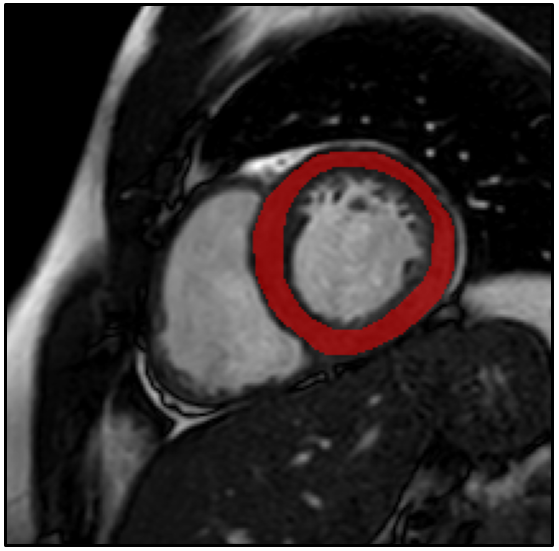
Figure
1. Example of a typical free-hand region
of interest drawn on a short axis bSSFP image, encompassing the left
ventricular myocardium.
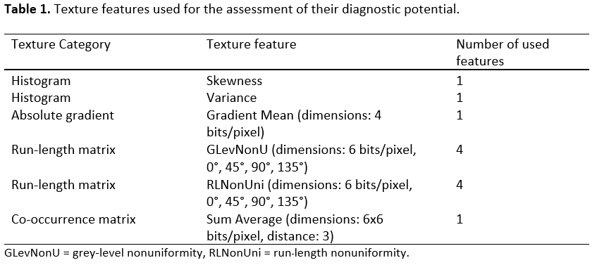
Table 1. Texture features used for
the assessment of their diagnostic potential.
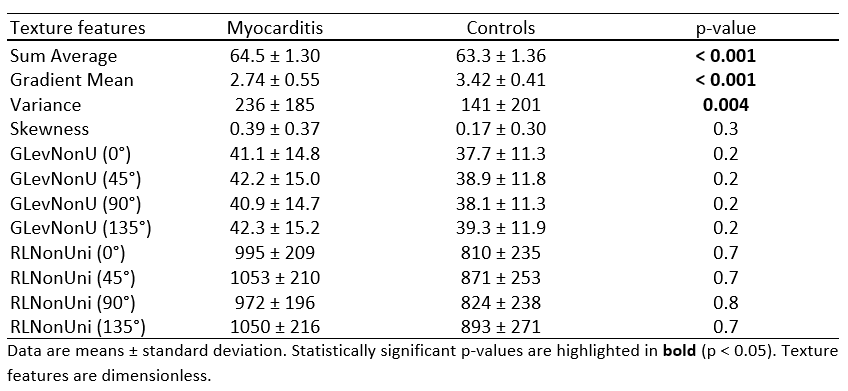
Table 2. Calculated means and standard deviations and estimated p-values of all
texture features in each group and technique.
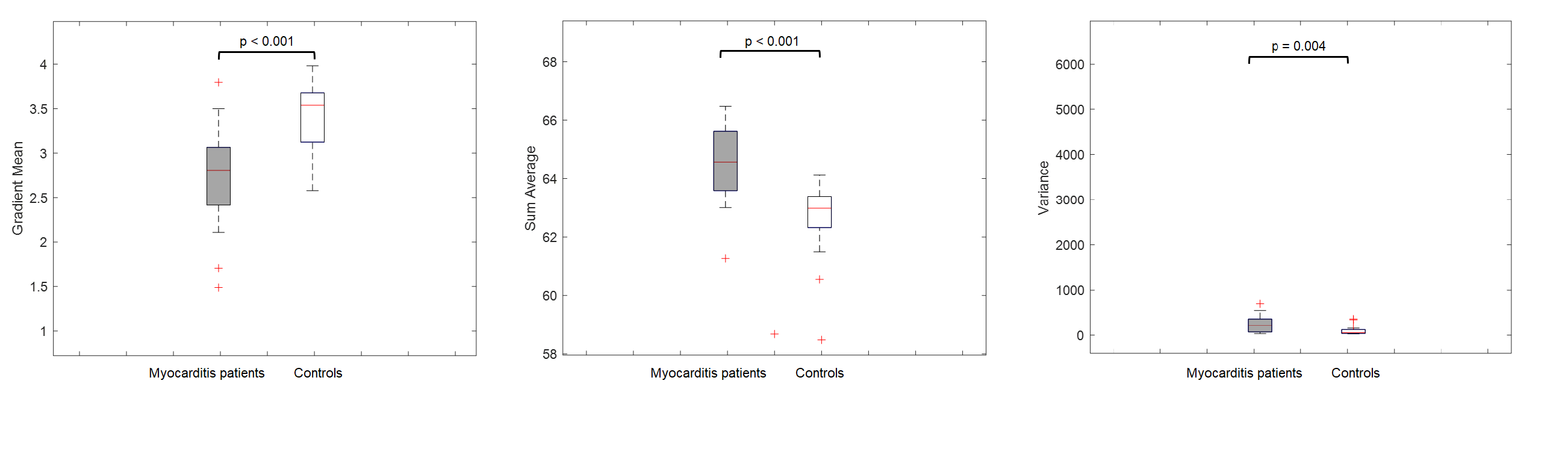
Figure 2. Box-Whisker
plots illustrating the differences for the significant texture features between
patients with myocarditis and controls on bSSFP images. The median is
represented by the centerline of the boxplot with upper and lower limits of
25th and 75th percentiles, respectively. The Whiskers extending from the boxes
indicates the most extreme values within 25th and 75th percentiles
±1.5*interquartile range; data points beyond the whiskers are displayed as +.
Texture features are dimensionless. bSSFP = balanced
steady-state-free-precession.