3603
Improvement of multi-echo gradient-spin-echo (mGraSE) myocardial T2 mapping utilizing Compressed SENSE reconstruction framework
Isao Shiina1, Michinobu Nagao2, Masami Yoneyama3, Yasuhiro Goto4, Kazuo Kodaira4, takumi ogawa4, Mamoru Takeyama4, Isao Tanaka4, and Shuji Sakai2
1Radiological Services, Tokyo Women's Medical University Hospital, tokyo, Japan, 2Department of Diagnostic Imaging and Nuclear Medicine, Tokyo Women's Medical University Hospital, Tokyo, Japan, 3Philips Electronics Japan, Tokyo, Japan, 4Department of Radiological Services, Tokyo Women's Medical University Hospital, Tokyo, Japan
1Radiological Services, Tokyo Women's Medical University Hospital, tokyo, Japan, 2Department of Diagnostic Imaging and Nuclear Medicine, Tokyo Women's Medical University Hospital, Tokyo, Japan, 3Philips Electronics Japan, Tokyo, Japan, 4Department of Radiological Services, Tokyo Women's Medical University Hospital, Tokyo, Japan
Synopsis
Myocardial T2 mapping using a multi-echo gradient-spin-echo (mGraSE) is widely used in clinical practice for quantitatively evaluating myocardial tissue properties with single breath-hold scan, but the limited scan time during the breath-hold period often results in poor signalto-noise ratio. Compressed SENSE has recently been developed to accelerate the acquisition time. Although C-SENSE is basically applied for non-EPI scans, mGraSE can also be applied the C-SENSE reconstruction framework. mGraSE myocardial T2 mapping with C-SENSE demonstrated improved image quality with higher image uniformity entire the shot-axis myocardium compared with conventional SENSE.
Introduction
Myocardial T2 mapping is useful in diagnosis of heart diseases, such as diffuse myocardial injury, mild myocardial fibrosis, and myocardial edema, because it allows quantitative evaluation [1] . A multi-echo gradient-spin-echo (mGraSE) sequence [Figure 1] is typically used for myocardial T2 mapping with single breath-hold scan [2,3]. However, the limited scan time during the breath-hold period often results in poor signal-to-noise ratio (SNR) and spatial resolution. One of the solution to shorten the scan time is to increase the parallel imaging (SENSE) reduction factor, but it often resulting in increasing the coil geometry-factor related noise and motion artifacts. Recently, a combination of parallel imaging and compressed sensing technique (Compressed SENSE, C-SENSE) has been developed to accelerate the acquisition time without increasing the image artifacts4,5. Although C-SENSE is basically applied for non-EPI scans, one study demonstrated that the C-SENSE reconstruction could clearly reduce noise-like artifacts and significantly improve the image quality of EPI based DWI without further optimization of EPI sampling scheme6. We hypothesized that mGraSE can also be applied the C-SENSE reconstruction framework as well as DW-EPI. The purpose of this study was to demonstrate the feasibility of a combination of mGraSE and C-SENSE for improving myocardial T2 mapping.Methods
A total of five volunteers were examined on a 3.0T system (Ingenia, Philips Healthcare). The study was approved by the local IRB, and written informed consent was obtained from all subjects. We compared the image quality of mGraSE myocardial T2 map with conventional SENSE and CSENSE. To quantitatively evaluate the image quality, region-of-interests (ROIs) were placed on myocardium with divided by 6 segments on the short axis of the left ventricle images for measuring the standard deviation (SD) of the T2 values, and these values were assessed by Wilcoxon signal-rank test. Imaging parameters for mGraSE myocardial T2 mapping were as follows: FOV=350mm, voxel size=2.0*2.0mm, slice thickness=8mm, flip angle=90, TR=1000ms, TE=8.8ms, NSA=1, SENSE=2.4 with acquisition time of14s, and C-SENSE=3 with acquisition time of 12s.Results and Discussion
Representative mGraSE myocardial T2 mapping with conventional SENSE and C-SENSE images are shown in Figure 2、3.mGraSE myocardial T2 mapping with C-SENSE showed improved image quality. myocardial T2 mapping with C-SENSE also indicated lower average SD entire the shot-axis myocardium, it suggests the image uniformity was improved compared with the conventional SENSE images.Conclusion
mGraSE myocardial T2 mapping with C-SENSE demonstrated improved image quality with higher image uniformity entire the shot-axis myocardium compared with conventional SENSE images. This might be useful for further accurate myocardial tissue characterization.Acknowledgements
No acknowledgement found.References
1.Hamlin SA, et.al. Mapping the future of cardiac MR imaging: case-based review of T1 and T2 mapping techniques.AE.Radiographics. 2014 Oct; 34(6):1594-611. 2. Sprinkart AM, et al. Gradient Spin Echo (GraSE) imaging for fast myocardial T2 mapping. J Cardiovasc Magn Reson 2015;17:12. 3. Fernández-Jiménez R, et al. Fast T2 gradient-spin-echo (T2-GraSE) mapping for myocardial edema quantification: first in vivo validation in a porcine model of ischemia/reperfusion. J Cardiovasc Magn Reson 2015;17:92. 4. Geerts-Ossevoort L, et al. Compressed SENSE Speed done right. Every time. The Netherlands: Philips Healthcare; 2018 Jan. Report No: 4522 991 31821. https://www.philips.de/content/dam/b2bhc/de/resourcecatalog/landingpages/ingeniaelition/White_Paper_Compressed_SENSE-opt.pdf 5. Mönch S, et al. Magnetic Resonance Imaging of the Brain Using Compressed Sensing - Quality Assessment in Daily Clinical Routine. Clin Neuroradiol. 2019 May 16. doi: 10.1007/s00062-019-00789-x. [Epub ahead of print] 6. Yoneyama, et al. Noise Reduction in Prostate Single-Shot DW-EPI utilizing Compressed SENSE Framework/ Proc. ISMRM. 2019:1634Figures
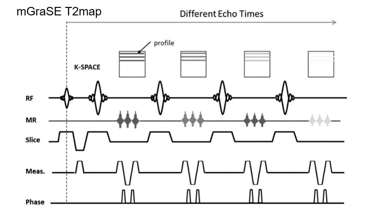
(Fig:1)Multishot Gradient Spin Echo (mGraSE) mapping sequences. mGraSE sequence an echo planar imaging (EPI) readout is interleaved
between each refocusing pulse, so that as many k-space lines are acquired as there are EPI factors, thus allowing shorter scan times.
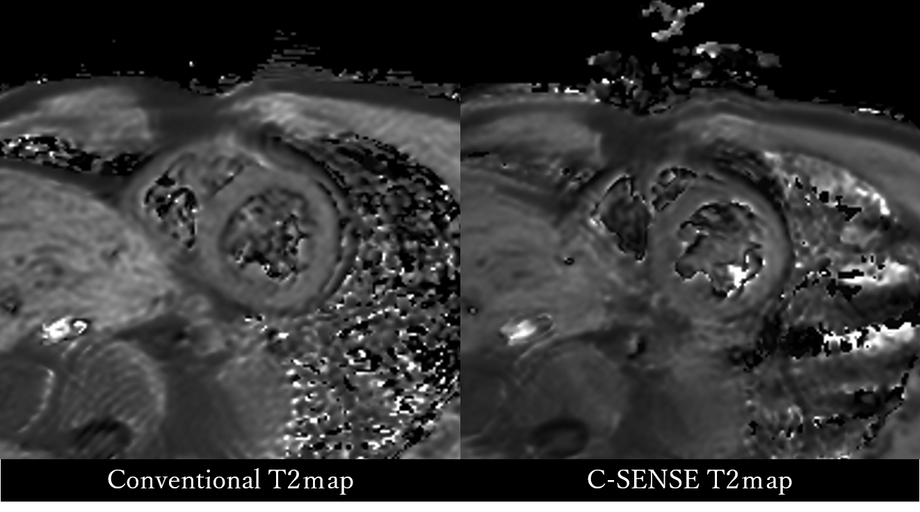
(Fig:2)A short-axis image of T2map. Comparison between conventional method and C-SENSE combined T2map
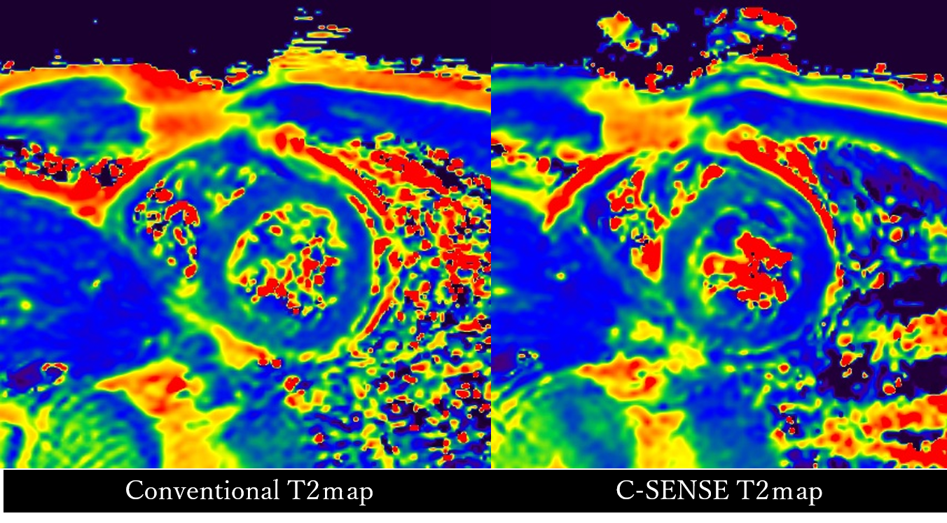
(Fig:3)A short-axis image of color T2map. Comparison between conventional method and C-SENSE combined color T2map
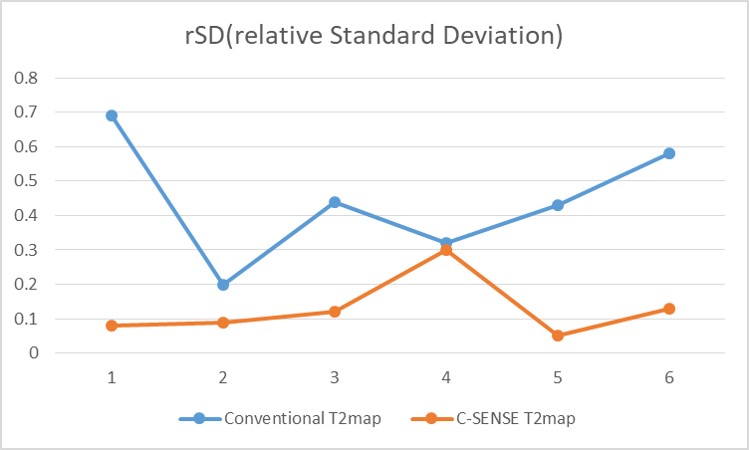
(Fig:4)Results of rSD in conventional T2map and C-SENSE T2map.respectively. C-SENSE T2map showed significantly higher signal uniformity
across all circumference of the myocardium compared to conventional T2 map.