3592
Hyperpolarized 13C-pyruvate MR imaging in rodent model of ventricular fibrillation cardiac arrest: A feasibility study1Emergency Medicine, Chonnam National University Hospital, Gwangju, Korea, Republic of, 2Biomedical Science, Chonnam National University, Gwangju, Korea, Republic of, 3Radiology, Chonnam National University, Gwangju, Korea, Republic of, 4Radiology, Chonnam National University Hospital, Gwangju, Korea, Republic of, 5Artificial Intelligence Convergence, Chonnam National University, Gwangju, Korea, Republic of
Synopsis
This study demonstrated that hyperpolarized 13C metabolic imaging can identify the metabolic changes within 1 hour of return of spontaneous circulation in rodent model of ventricular fibrillation-induced cardiac arrest. Brain histology performed at 72 hours after ROSC showed marked difference in the levels of neuronal death and reactive astrocyte change between the cardiac arrest model and sham control. The results from this study suggest that this new metabolic imaging technique may provide a unique and noninvasive imaging biomarker for assessing functional changes at early stage of post ROSC and warrant further investigation.
Introduction
Despite advances in cardiopulmonary resuscitation (CPR) and post-cardiac arrest (CA) care, only about 8.4% of CA survivors are discharged with a good neurologic outcome [1]. Although the identification of the sign of neurologic abnormality is a fundamental base of post-CA care for further supportive interventions, noninvasive, early and accurate measurement of functional changes in comatose CA survivors remains challenging despite various advanced imaging techniques. The purpose of this study was to explore the feasibility of HP 13C metabolic imaging in assessing metabolic changes following heart arrest. We hypothesized that hyperpolarized 13C MR with pyruvate can easily identify the metabolic changes within 1 hour of return of spontaneous circulation (ROSC) in the rodent model of ventricular fibrillation-induced CA (VFCA).Methods and Materials
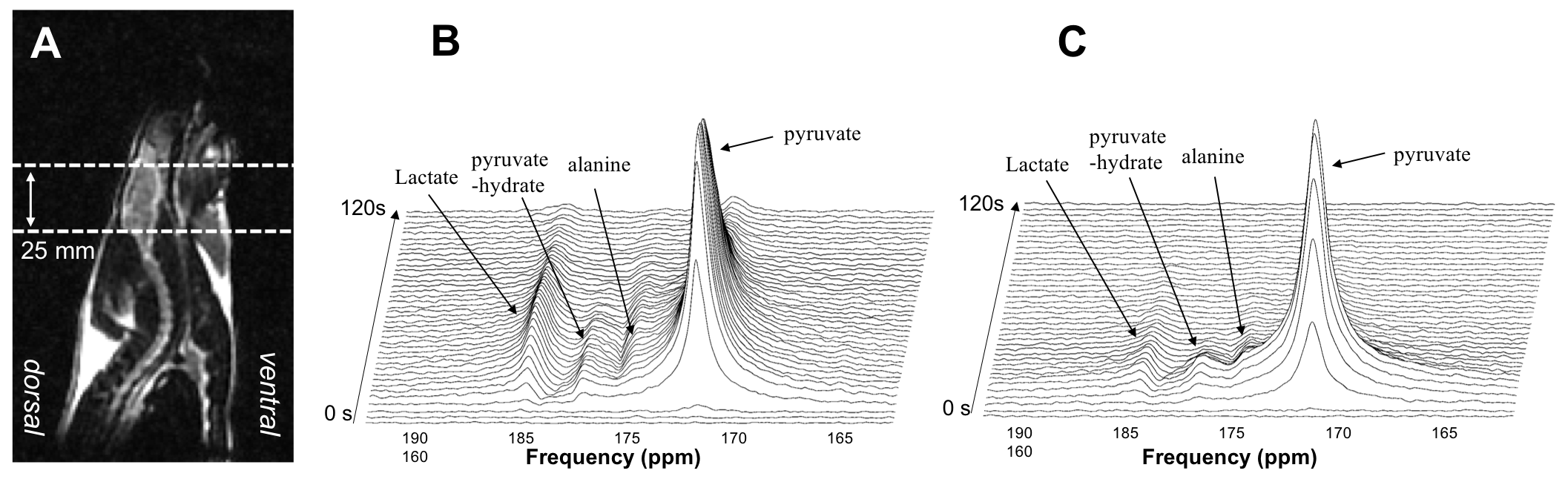
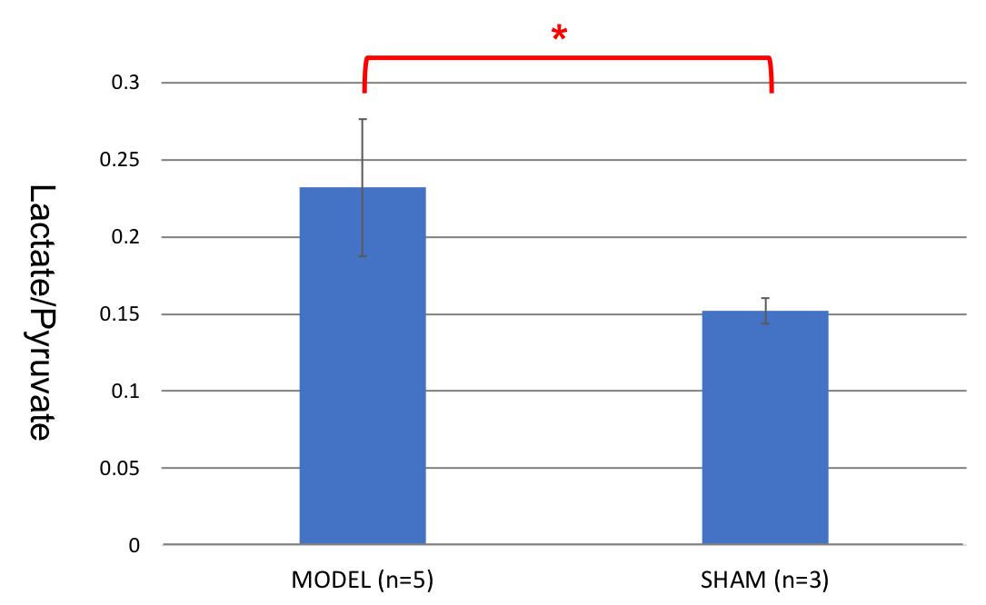
Eight male Sprague Dawley rats (5 CA models and 3 sham control), 9−10 weeks old weighing between 300g and 350g were included in this study. Under isoflurane anesthesia, oral intubation and mechanical ventilation were performed. End-tidal CO2 (EtCO2), rectal temperature, and blood pressure were continuously monitored. Anesthesia was discontinued 1.5min before inducing CA. VFCA was induced by trans-esophageal electrical stimulation (15V−60Hz alternating current for 1.5min). CA was confirmed by a drop in mean arterial blood pressure (MAP) and loss of pulsatility in the femoral arterial line. After 7min of untreated VFCA, resuscitation was started with metronome guided manual chest compressions (250/min) and mechanical ventilation. At 2 minutes of CPR, rats were defibrillated (2 times with 5 J, biphasic) and defibrillation attempts repeated every 2min. Epinephrine (20ug/kg) and bicarbonate(1mEq/kg) were given iv 30s prior to the start of CPR. Epinephrine alone at 10ug/kg was administered 90s after the start of CPR and repeated every 2min during CPR. If ROSC was not achieved within 10min from chest compression, the resuscitation was terminated. Upon achieving spontaneous circulation that defined mean arterial pressure above 50mmHg for 5min, animals were observed for 10 minutes. After additional 10 minutes stabilizing period, catheters were removed, surgical cut-downs repaired, and mechanical ventilation weaned off.The HP 13C metabolic imaging data were obtained within 1 hour of return of ROSC using a GE clinical 3T scanner with a custom 13C/1H dual rat coil. 32mL of [1-13C]pyruvate was polarized using a HyperSense DNP polarizer [2]. Slice-localized 13C MR spectroscopy (MRS) data (TE/TR=35/3000ms, flip angle=20, 3s temporal resolution, 60 total time points) were acquired from a 25mm slab centered in brain with an injection of dissolved 3mL HP 13C1-pyruvate solution (100 mM and pH of 7.5) through tail vein. The ratio of lactate over pyruvate (Lac/Pyr) was calculated from the temporally summed magnitude spectra and compared between the CA models and sham controls using a 2-tailed t-test.
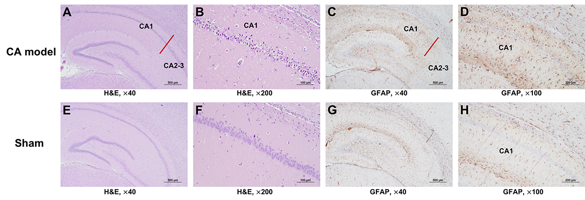
At 72 hours post-CA, the brains of rats were harvested and the following histopathology and immunohistochemistry stains were performed: hematoxylin&eosin (H&E) and glial fibrillary acidic protein (GFAP).
Results and Discussion
The use of hyperpolarized 13C1-pyruvate provided sufficient signal to detect its transfer of the 13C label to lactate and alanine. Figure 1 shows representative 13C dynamic spectra from a 25 mm slice of rat brain from a CA model (Fig 1B) and a sham control (Fig 1C). Typically, the 13C1-pyruvate signal (173 ppm) reached its maximum at approximately 15 seconds after the start of hyperpolarized pyruvate injection and was followed by the 13C1-lactate signal maximum (185 ppm) at approximately 20 seconds following pyruvate injection.Quantification of the HP 13C MRS data is depicted in Figure 2. Lac/Pyr of CA models was significantly higher than that of sham controls, indicating a cellular metabolic change occurring in the brain of CA animals at an early stage of spontaneous circulation.
The histopathological and immunohistochemical analysis revealed marked differences between the brains of the cardiac arrest model and sham control. Figure 3 shows H&E and GFAP stained slices with various magnifications from region CA1 and CA2-3 of the brains from a cardiac arrest model (top row) and sham control (bottom row). Region CA1 of the cardiac arrest model, which is known to be an area that is affected by ischemic injury, exhibited a large amount of neuronal death (Fig 3A and 3B), while the same region of the sham control did not exhibit noticeable neuronal damage (Fig 3E and 3F). Region CA1 of the cardiac arrest model demonstrated a relatively higher amount of GFAP staining compared to region CA2-3 (Fig 3C) with a large level of reactive astrocyte change (Fig 3D). In contrast, the sham control demonstrated a comparable level of GFAP staining between region CA1 and CA2-3 with a relatively small amount of positive staining compared to the cardiac arrest model.
Conclusion
This study demonstrated that hyperpolarized 13C metabolic imaging can identify the metabolic changes within 1 hour of the return of ROSC in the rodent model of ventricular fibrillation-induced cardiac arrest. Brain histology performed at 72 hours after ROSC showed a marked difference in the levels of neuronal death and reactive astrocyte change between the cardiac arrest model and sham control. The results from this study suggest that this new metabolic imaging technique may provide a unique and noninvasive imaging biomarker for assessing functional changes at an early stage of post-ROSC and warrant further investigation.Acknowledgements
This study was supported by the Ministry of Education, Republic of Korea (2019R1I1A3A01059201) and the Korea Health Technology R&D Project through the Korea Health Industry Development Institute (KHIDI), funded by the Ministry of Health & Welfare, Republic of Korea (HR20C0021).References
1. 2017 Cardiac Arrest Registry to Enhance Survival (CARES). https://mycares.net.
2. Ardenkjaer-Larsen JH, Fridlund B, Gram A, et al. Increase in signal-to-noise ratio of > 10,000 times in liquid-state NMR. Proc Natl Acad Sci USA. 2003;100(18):10158-63.
Figures