3571
Template-based bias field correction of Hyperpolarized 129Xe Gas Ventilation MRI1Medical Physics, Duke University, Durham, NC, United States, 2Radiology, Duke University, Durham, NC, United States, 3Biomedical Engineering, Duke University, Durham, NC, United States
Synopsis
Accurately correcting hyperpolarized 129Xe ventilation MRI for coil-induced bias field remains the most significant obstacle to precise, repeatable quantitative image analysis. Estimates using B1 maps from RF-depletion in radially acquired images may provide an improvement on the standard N4ITK solution, which recent works suggests may perform an overly aggressive correction. Here, we develop a template-based approach to bias field correction using B1 maps derived from multiple subjects. This paradigm is then evaluated by applying it to a set of test images reflective of a range of disease types and levels of ventilation obstruction.
Introduction
Hyperpolarized 129Xe magnetic resonance imaging (129Xe MRI) enables sensitive regional assessment of ventilation across numerous pulmonary diseases and visualizes regional therapy response in both adult and pediatric cohorts1,2. 129Xe ventilation MRI can be quantified by numerous approaches such as linear binning and k-means classification of the signal distribution3. However, an ongoing challenge for these methods is to identify and correct for signal variation caused by coil-induced B1-inhomogeneity, while still retaining the heterogeneity attributable to underlying physiology. Previous work has shown that the most common solution N4ITK4 provides an overly aggressive bias field correction5. This work also demonstrated an approach based on RF-depletion mapping5 that directly calculates B1 using the relative signal decay of two temporal-subdivisions of a 3D radial acquisition6. However, RF-depletion mapping is applicable only to center-out 3D-radial acquisitions, which are not yet as widely adopted as Cartesian imaging. To address this issue, we introduce and demonstrate a template-based approach7 to correct for B1-inhomogeneity for a given coil configuration that can be used with any acquisition strategy.Methods
1) ImagingData were selected for analysis from 15 subjects who had undergone radial 129Xe ventilation MRI in the supine position at 3T (Siemens Magnetom Trio Scanner VB19) using the following parameters: views=3600; samples/view=128; TR/TE=4.5/0.45ms; flip angle=1.5; FOV=40cm. All 3D volumes were acquired using a randomized 3D Halton spiral radial sequence and reconstructed to a size of 128x128x128 with a constant kernel sharpness of 0.328. Images were bias field corrected using both RF-depletion mapping5 and N4ITK4. Both methods further employ a B-spline model to create a smoothly varying bias field across the whole volume. Segmentation of the thoracic cavity was conducted by an expert reader.
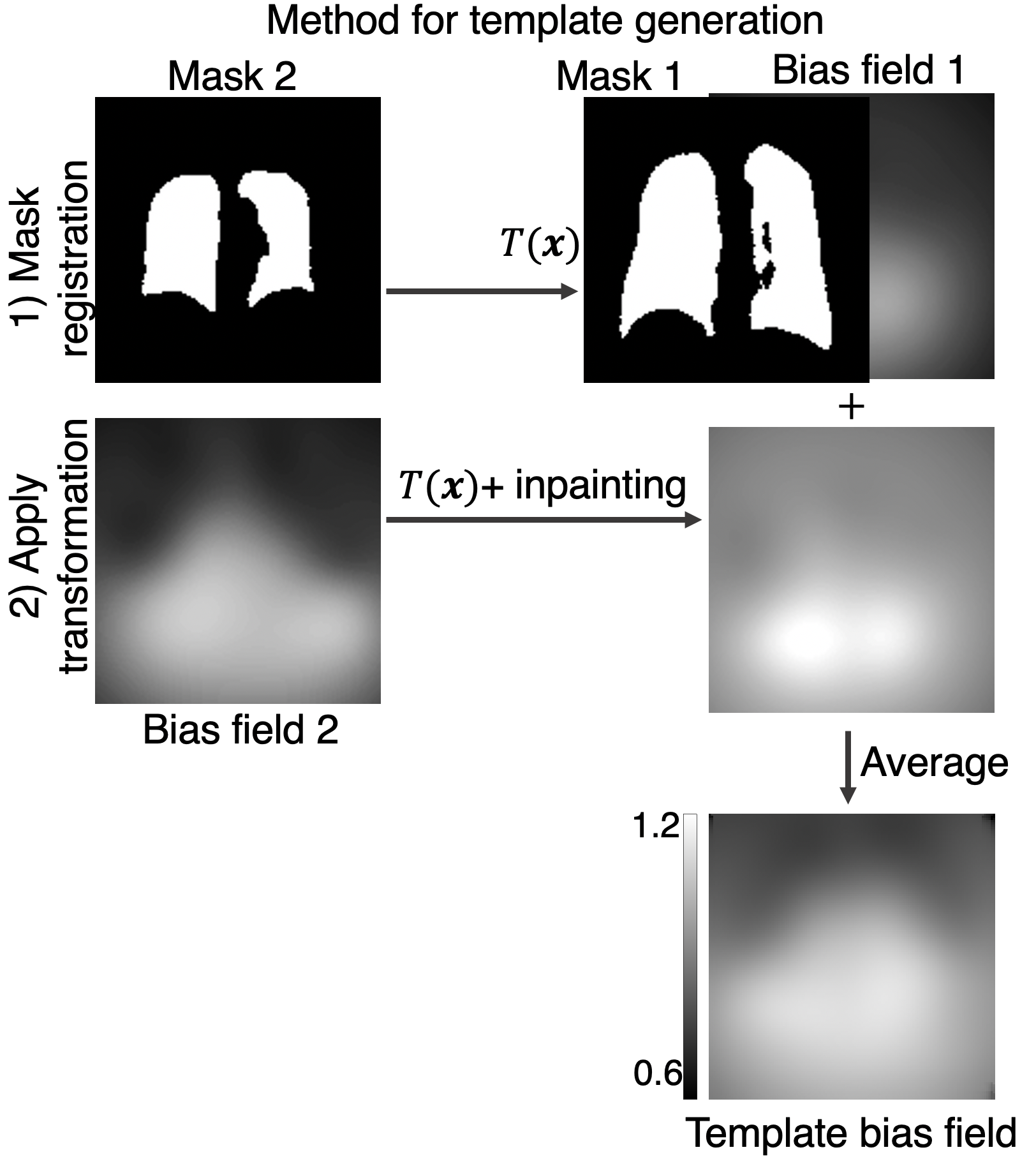
2) Template Generation
The bias field template was generated from 129Xe ventilation images from a subset of 10 selected subjects (Figure 1). For each subject, the thoracic cavity masks were registered to a common coordinate system. These same transformations were applied to the subject’s RF-depletion-derived bias field. Dark areas at the boundary caused by the transformed volume moving partially out of the FOV were inpainted9 and then all 10 bias fields were averaged.
3) Template Correction
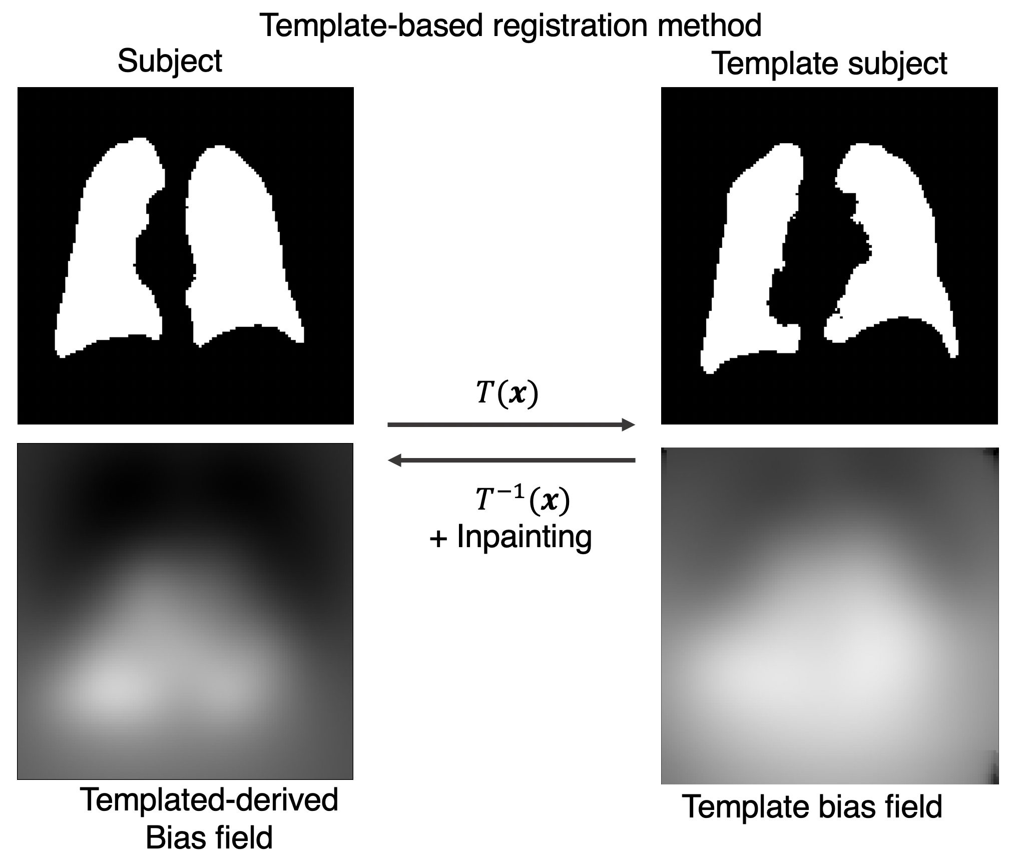
The bias field template $$$b_{template}(\textbf{x})$$$ was associated with a thoracic cavity mask given by $$$m_{template}(\textbf{x})$$$ that was used to register the template to the uncorrected scan of interest. The bias field is modeled as a smoothly varying multiplicative field such that local registration issues will not significantly affect bias field estimate. The subject and bias field template masks are mapped to one another via registration by
$$m_{template}(\textbf{x}) = m_{subject}(\textbf{x})$$
where $$$T(\textbf{x})$$$ is the mapping function. Its inverse permits the bias field of the subject image to be derived from the template as
$$b_{subject}(\textbf{x}) = b_{template}(\textbf{x})$$
With this estimate of the bias field, the subject’s uncorrected ventilation $$$v(\textbf{x})$$$ can simply be corrected by
$$v_{corrected}(\textbf{x}) = v(\textbf{x})/b_{subject}(\textbf{x})$$
where $$$v_{corrected}(\textbf{x})$$$ is the bias-field correct image, as depicted in Figure 2. To test this approach, the template bias field was then registered to each of the 5 remaining cases and used to bias field correct them. For each form of bias correction (template, RF-depletion, and N4ITK), we compare the rescaled10 corrected ventilation distribution.
Results
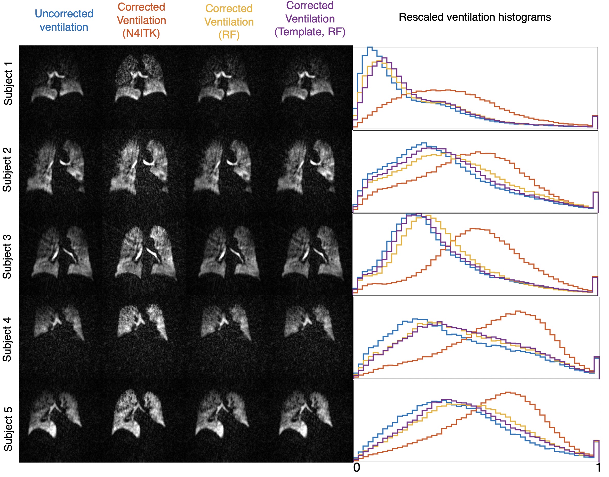
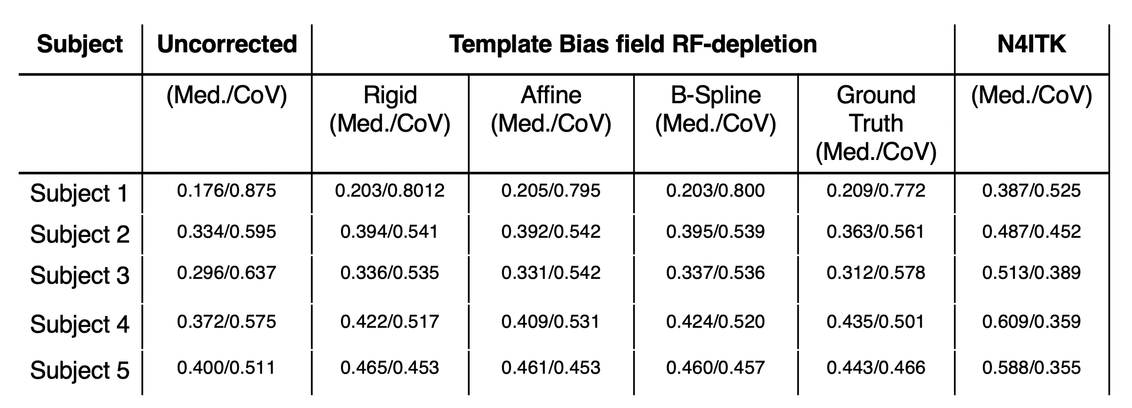
Figure 3 compares the visual effects of template-based bias field correction using rigid transformation to that of N4ITK and RF-depletion mapping bias field correction. For images corrected using the RF-depletion derived bias field template, the corrected ventilation distribution is in good visual agreement with images corrected using the known individual subject’s RF-depletion bias field. Moreover, for images corrected using N4ITK, corrections are significantly more aggressive, as indicated by the skewing of the ventilation distribution histograms.These results are further quantified in the table (Figure 4), which characterizes the effect of bias field correction using the mean and the coefficient of variation inside the thoracic cavity to characterize the effect of bias field correction. The median values reflect how much the ventilation distributed shifted and the coefficient of variation is used to compare the signal uniformity inside the thoracic cavity. Notably, the method of the type transformation (either rigid, affine, or b-spline transformation) does not significantly affect the median and coefficient of variation of the ventilation distribution.
Discussion
Bias field correction using generated template bias fields could serve as a simpler, more generally applicable alternative to current efforts to generate a bias field correction for each individual image. This approach is less susceptible to applying overly harsh corrections and appears more robust against severe defects and physiological gradients. Although demonstrated for only a single coil configuration with 10 subjects, this method can extend to having more templates to account for other coil configurations or other template bias field estimation methods. This method offers the unique benefit of avoiding direct RF-depletion mapping for each individual image while also avoiding the overly aggressive and naïve N4ITK correction. Moreover, successful correction of the subject images in this study indicates that signal inhomogeneity due to bias field is indeed repeatable for a given coil configuration. This approach to bias field correction that incorporate these knowledge priors while avoiding the need for individual B1-mapping may provide a valuable improvement to 129Xe MRI image quantification.Acknowledgements
R01HL105643, R01HL12677, NSF GRFP DGE-1644868References
1. Wang, Z. et al. Diverse cardiopulmonary diseases are associated with distinct xenon magnetic resonance imaging signatures. Eur. Respir. J. 54, (2019).
2. Thomen, R. P. et al. Hyperpolarized 129Xe for investigation of mild cystic fibrosis lung disease in pediatric patients. J. Cyst. Fibros. 16, 275–282 (2017).
3. He, M. et al. A Comparison of Two Hyperpolarized 129Xe MRI Ventilation Quantification Pipelines: The Effect of Signal to Noise Ratio. Acad. Radiol. 26, 949–959 (2019).
4. Tustison, N. J., Cook, P. A. & Gee, J. C. N4ITK: Improved N3 Bias Correction. 29, 1310–1320 (2011). 5. Lu, J. et al. Bias Field Correction in Hyperpolarized 129 Xe Gas Ventilation MRI. ISMRM (2020).
6. Niedbalski, P. J. et al. Mapping and correcting hyperpolarized magnetization decay with radial keyhole imaging. Magn. Reson. Med. 82, 367–376 (2019).
7. Studholme, C. et al. Accurate Template-Based Correction of Brain MRI Intensity Distortion with Application to Dementia and Aging. IEEE Trans. Med. Imaging 23, 99–110 (2004).
8. Robertson, S. H. et al. Optimizing 3D noncartesian gridding reconstruction for hyperpolarized 129 Xe MRI-focus on preclinical applications. Concepts Magn. Reson. Part A 44, 190–202 (2015).
9. Bertalmío, M., Bertozzi, A. L. & Sapiro, G. Navier-Stokes, fluid dynamics, and image and video inpainting. Proc. IEEE Comput. Soc. Conf. Comput. Vis. Pattern Recognit. 1, 355–362 (2001).
10. He, M., Driehuys, B., Que, L. & Yuh-Chin, H. Using Hyperpolarized 129Xe MRI to Quantify the Pulmonary Ventilation Distribution. Acad. Radiol. 270–274 (2016).
Figures