3569
Detection of pulmonary abnormalities in a rabbit thoracic insufficiency syndrome model using hyperpolarized xenon-129 MRI1University of Pennsylvania, Philadelphia, PA, United States, 2School of Veterinary Medicine, University of Pennsylvania, Kennett Square, PA, United States, 3Children's Hospital of Philadelphia, Philadelphia, PA, United States, 4Boston Children's Hospital, Boston, MA, United States
Synopsis
Thoracic insufficiency syndrome (TIS) progresses to the development of restrictive lung disease and is commonly treated through surgical intervention. In this work, we used a rib-tether rabbit model to investigate the sensitivity of dynamic 1D simultaneous dissolved- and gas-phase hyperpolarized xenon-129 MRI imaging to pulmonary abnormalities secondary to TIS. We found asymmetric lung ventilation patterns and increases in alveolar septal wall thickness in both lungs of a rib-tethered rabbit compared to an age-matched control animal. These findings could help identify the optimal timepoint at which to conduct chest expansion surgery so as to maximize the resulting improvements in lung maturation.
Purpose
Thoracic insufficiency syndrome (TIS)1 is associated with multiple pediatric disorders including cerebral palsy, spinomuscular atrophy, and congenital malformations of the spine2,3. TIS typically progresses to the development of restrictive lung disease4,5, putting patients at a significantly increased risk of pulmonary hypertension and chronic respiratory failure6-8. While surgical management of TIS can substantially ameliorate its negative impact on respiratory function and mortality, the optimal timing of intervention remains unclear. Early surgical intervention has the potential to rescue alveolar formation9, but must be justified against the increased risk of surgery at a young age. To develop tools for assessing the effect of different treatment approaches on lung function, we performed hyperpolarized xenon-129 (HXe) MRI studies using dynamic simultaneous gas-phase (GP) and dissolved-phase (DP) 1D acquisitions to study the effect of chest restriction in a rib-tether rabbit model.Methods
Rib-tethering was performed in New Zealand White rabbits (3-6 weeks of age; 0.7-1.3 kg body weight). A longitudinal skin incision was made along the dorsal angle of the right hemithorax along ribs 1 through 9, and the dissection was deepened to the intercostal muscles. The periosteum of eight ribs (2-9) was incised and the rib elevated from its periosteal bed. A figure-of 8 ligature was used to tether the ribs together, consequently constricting growth and inducing acute thoracic insufficiency of the right hemithorax. At 28 weeks of age, the first rabbit of this cohort and an age-matched healthy control animal were orotracheally intubated and maintained on a Propofol at a continuous infusion rate (20-80 mg/kg/hr). Animals were ventilated with room air until imaging began, at which point the gas mix was switched to 20% oxygen and 80% HXe for 15 breaths (6 ml/kg tidal volume). All studies were approved by the Institutional Animal Care and Use Committee.MR imaging was conducted using a 1D-projection gradient-echo sequence with left-to-right frequency encoding that employed a non-selective 700-ms Gaussian RF excitation pulse centered at the DP resonance, 3,530 Hz downfield from the GP resonance. Taking advantage of the large frequency difference between the two phases, combined with a sufficiently small acquisition bandwidth, HXe in the pulmonary air spaces and dissolved in the lung tissue were imaged simultaneously, side-by-side.10-12 The following sequence parameters were used: matrix size 1920×80; TE 2.6 ms; FOV 220 mm; receiver bandwidth 120 Hz/pixel; flip angle 7°, TR 10 ms (TR90°,equiv 1.3 s12). During ventilation with xenon gas, the DP magnetization was saturated every 500 ms with 3 consecutive 3-ms frequency-selective Gaussian RF pulses centered at 200 ppm and separated by 1.2 ms spoiler gradients. GP and DP signals were analyzed for left and right lungs in aggregate. In order to fit the DP signal at 4 different lung inflation levels to the analytical uptake model of Patz et al.13, the DP signal was divided into four bins based on the GP signal level. All MR studies were performed at 1.5T (Avanto; Siemens) using a custom xenon-129 transmit/receive birdcage coil (Stark Contrast, Erlangen, Germany). Enriched xenon gas (87% xenon-129) was polarized using a prototype commercial system (XeBox-E10, Xemed LLC, Durham, NH).
Results and Discussion
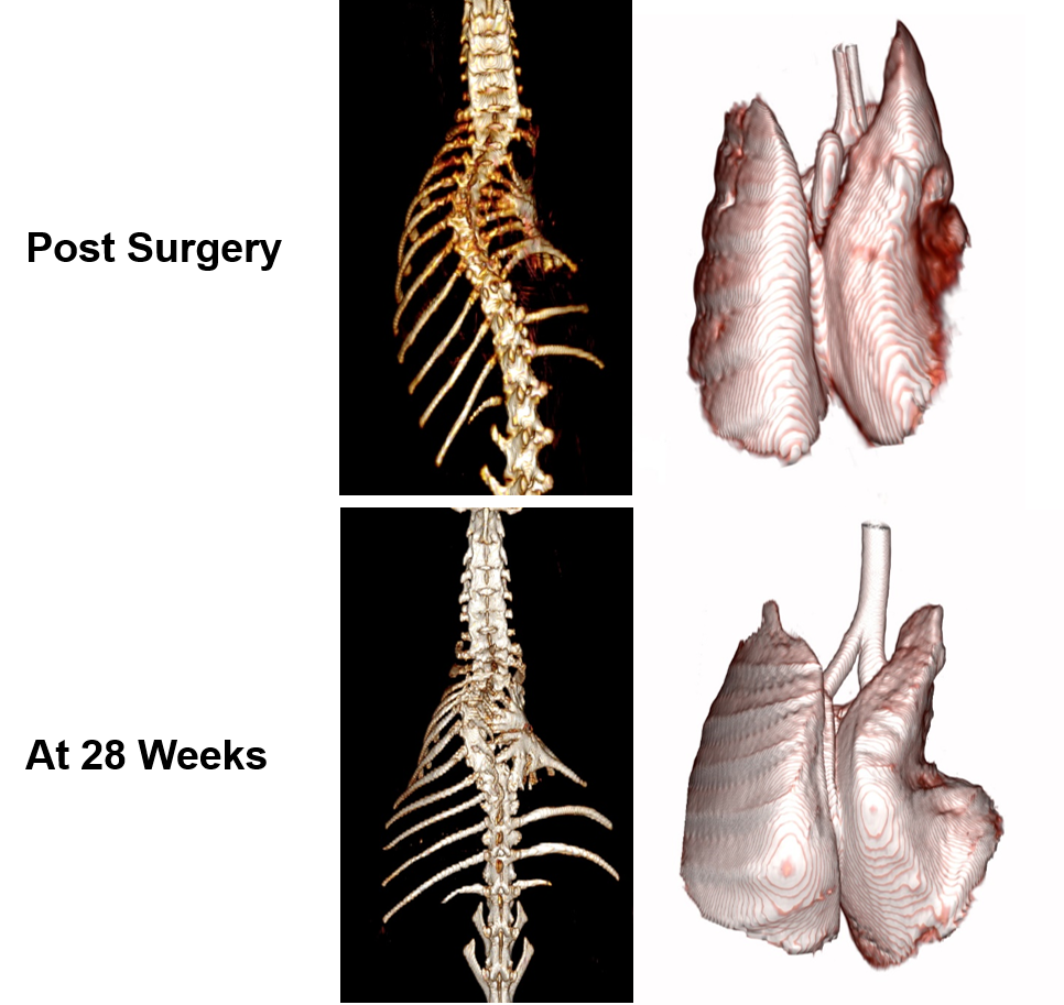
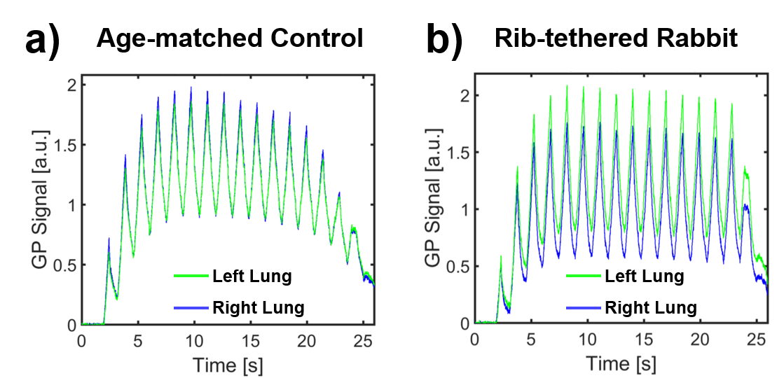
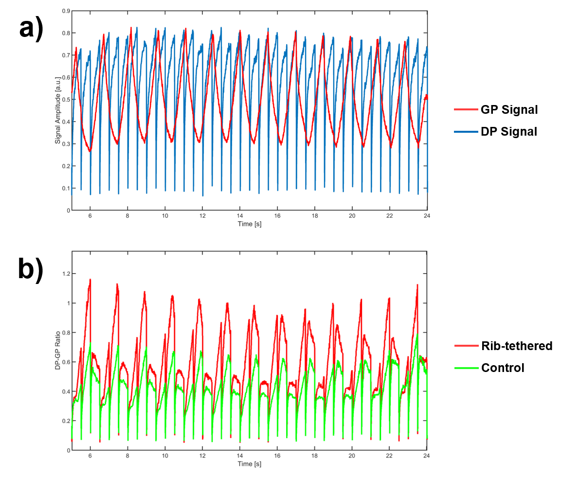
Figure 1 depicts CT-based surface renderings of the spine and lung in the rib-tethered rabbit after surgery at approximately 6 and 28 weeks of age, respectively. The GP signal for the healthy control animal (Figure 2a) indicates symmetric lung inflation during mechanical ventilation. In the rib-tethered rabbit (Figure 2b), ventilation in the ipsilateral right lung is substantially lower than in the contralateral left lung, an effect that is likely underestimated by positive-pressure mechanical ventilation. Figure 3a illustrates a representative plot of the dynamic total GP and DP signals during the multi-breath acquisition. The DP signal is saturated every 500 ms and its recovery sampled with low flip angle RF pulses. The associated dynamic DP-GP ratio for the rib-tethered and control animals are shown in Figure 3b. Throughout the respiratory cycle, the DP-GP ratio in the rib-tethered rabbit is generally 20-80% higher than in the control rabbit. Interestingly, the maximum ratio difference occurs at end expiration, and thus cannot be attributed to differences in lung compliance. Further, the septal wall thickness for all ventilation levels in the control animal was 12.5 μm (left lung 12.5 +/- 1.2 μm, right lung 12.5 +/- 1.3 μm), but 18.0 +/- 2.1 μm in the ipsilateral and 14.7 +/- 1.0 μm in the contralateral lung of the rib-tethered rabbit, which might be an indication of incomplete lung maturation.Conclusion
Using dynamic 1D HXe MRI measurements, we detected abnormal lung physiology in a surgical TIS rabbit model. Our findings of asymmetric breathing mechanics as well as increased DP-GP ratios and septal wall thicknesses even in the contralateral lung support the hypothesis that HXe MRI is sensitive to TIS-related pulmonary abnormalities but need to be confirmed in larger cohorts. Future research will also explore at which growth stage surgical chest expansion still promises adequate lung maturation while minimizing the number of repeat surgeries during body growth to adult size. Since all acquisitions can be performed during free breathing, these imaging techniques are highly translatable to non-cooperative subjects such as young children.Acknowledgements
Supported by the Wyss/Campbell Center for Thoracic Insufficiency Syndrome.References
[1] Campbell RM, Smith MD, Mayes TC, Mangos JA, Willey-Courand DB, Kose N, et al. The characteristics of thoracic insufficiency syndrome associated with fused ribs and congenital scoliosis. J Bone Joint Surg Am 2003;85-A:399–408.
[2] Campbell RM, Smith MD. Thoracic insufficiency syndrome and exotic scoliosis. J Bone Joint Surg Am 2007;89 Suppl 1:108–22.
[3] Mayer O, Campbell R, Cahill P, Redding G. Thoracic Insufficiency Syndrome. Curr Probl Pediatr Adolesc Health Care 2016;46:72–97.
[4] Tsiligiannis T, Grivas T. Pulmonary function in children with idiopathic scoliosis. Scoliosis 2012;7:7.
[5] Karol LA, Johnston C, Mladenov K, Schochet P, Walters P, Browne RH. Pulmonary function following early thoracic fusion in non-neuromuscular scoliosis. J Bone Joint Surg Am 2008;90:1272–81.
[6] Pehrsson K, Larsson S, Oden A, Nachemson A. Long-term follow-up of patients with untreated scoliosis. A study of mortality, causes of death, and symptoms. Spine 1992;17:1091–6.
[7] Koumbourlis AC. Scoliosis and the respiratory system. Paediatr Respir Rev 2006;7:152–60.
[8] Kafer ER. Respiratory and cardiovascular functions in scoliosis. Bull Eur Physiopathol Respir 1977;13:299–321.
[9] Olson JC, Takahashi A, Glotzbecker MP, Snyder BD. Extent of Spine Deformity Predicts Lung Growth and Function in Rabbit Model of Early Onset Scoliosis. PLoS ONE 2015;10:e0136941.
[10] Mugler et al. Simultaneous magnetic resonance imaging of ventilation distribution and gas uptake in the human lung using hyperpolarized xenon-129. Proc Natl Acad Sci USA 2010;107(50):21707-21712.
[11] Ruppert et al. Assessment of Pulmonary Gas Transport in Rabbits Using Hyperpolarized Xenon-129 Magnetic Resonance Imaging. Scientific reports 8, 7310, doi:10.1038/s41598-018-25713-0 (2018).
[12] Ruppert et al. Assessment of flip angle-TR equivalence for standardized dissolved-phase imaging of the lung with hyperpolarized 129Xe MRI. MRM 2019; 81(3):1784-1794.
[13] Patz et al. Diffusion of hyperpolarized 129Xe in the lung: a simplified model of 129Xe septal uptake and experimental results. New J Physics 2011;13:015009.
Figures