3563
A 129Xe/1H Switched Frequency High Pass Birdcage Coil for Hyperpolarized 129Xe Gas Lung Imaging in Neonates at 1.5 T1Imaging Research Center/Radiology, Cincinnati Children's Hospital Medical Center, Cincinnati, OH, United States, 2Center for Pulmonary Imaging Research/Radiology, Cincinnati Children's Hospital Medical Center, Cincinnati, OH, United States, 3POLARIS, Imaging Sciences, Dept of Infection, Immunity & Cardiovascular Disease, University of Sheffield, Sheffield, United Kingdom
Synopsis
Production of hyperpolarized 129Xe gas has opened new frontiers for imaging lung anatomy and function using MRI. Our institution has the goal of using hyperpolarized 129Xe gas for MR imaging of neonatal lungs at 1.5 T. To realize this goal, it is essential that the RF coil used for imaging operate at both the 129Xe and 1H frequencies. This report provides fabrication details of a novel 129Xe/1H switched frequency transmit/receive 16 rung high-pass birdcage RF coil. Images collected with hyperpolarized 129Xe gas and water in phantoms demonstrate that the coil provides excellent image quality at both frequencies.
Introduction:
The production of hyperpolarized 129Xe and 3He gases has opened new and exciting possibilities for imaging lung anatomy and function using MRI 1-4. Our institution has the goal of using hyperpolarized 129Xe gas for MR imaging of neonatal lungs using a dedicated multinuclear Neonatal Intensive Care Unit (NICU)-housed 1.5T MR scanner developed at our institution 5-6. While hyperpolarized 129Xe gas imaging provides unique information into lung function and microstructure, complementary 1H MRI is needed to provide anatomic references, structural images of lung parenchyma, and B0 and B1 maps. Further, minimal disruption of neonatal subjects is advantageous since this population is delicate. With these aims and requirements in mind, it is essential that the MRI coil used for imaging can operate at both 129Xe and 1H frequencies without a signal-to-noise penalty at either frequency and without the requirement of moving the patient between scans for coil changes.We have implemented a novel design for a transmit/receive 16 rung high-pass birdcage RF coil that can be used to collect high quality images at both 129Xe and 1H frequencies (17.76 MHz and 63.86 MHz respectively at 1.5 T). Advantages of this coil design include: 1) quadrature excitation and reception at both frequencies, 2) good SNR and B1 homogeneity at both frequencies, and 3) 10 - 20 second switching times between 129Xe and 1H operation without patient disruption. This report provides details of the coil fabrication. Images of hyperpolarized 129Xe gas and 1H in phantoms demonstrate the image quality obtained with the coil.
Materials and Methods:
A 129Xe/1H switched frequency 16-rung high pass birdcage coil was constructed on a G10 (garolite) cylinder. The coil has an inner diameter of 178 mm (7.0 in) which will accommodate more than 90% of the neonates in our NICU. The coil rung length is 140 mm (5.5 in) long, which is adequate to cover the neonatal lung field. The outer shell of the coil was constructed from a second G10 cylinder that also supports an integrated RF shield.Figure 1 shows the coil without its outer cylinder. Thirty-two (32) beryllium-copper (BeCu) contacts (red arrow) are attached to FR4 fiberglass slats (green arrow). The slats are connected in groups of four to eight polycarbonate pieces (white arrow). Each set of four contacts is positioned by a rod (blue arrow) connected to the polycarbonate pieces. When the rods are inserted, the BeCu contacts bridge (short) gaps (yellow arrow) on the coil printed circuit board (PCB) placing 32 banks of additional capacitors (orange arrows) in parallel with the birdcage’s main capacitors (purple arrows). The added capacitance shifts the resonance frequency of the coil from 1H down to 129Xe. When the rods are retracted, the gaps in the PCB are no longer bridged and the added capacitor banks are removed from the circuit. This shifts the resonance frequency of the coil back to the 1H frequency. Shielded cable baluns (not shown) on the I and Q channels provide 5.6 K ohms isolation impedance.
Results and Outlook:
129Xe and 1H imaging of phantoms was performed using the switched frequency high pass birdcage coil on our NICU 1.5T MRI scanner.1H imaging: Figure 2 shows six axial images (of 256 total images) of a water filled cylindrical phantom. The images were acquired using a 3D radial ultrashort echo time (UTE) spoiled gradient echo imaging sequence7. Acquisition parameters: ~200,000 radial projections; TE/TR = 0.2/5 msec; FA = 5 degrees; FOV = 18 cm; reconstructed to a matrix size of = 256 x 256 x 256, resolution = 0.7 mm3. The average SNR for the six slices is ~33.
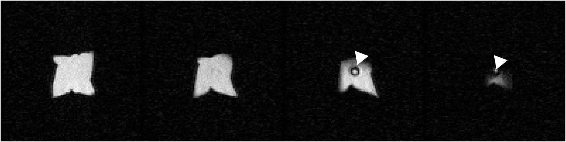
129Xe imaging: Phantom description: Tedlar® bag of volume ~300 ml; xenon gas volume 200 ml (with ~85% 129Xe). The 129Xe gas was generated at a polarization of ~30%. Figure 3 shows four coronal 129Xe images obtained using a 2D slice-selective spoiled gradient echo (SPGR) sequence. Acquisition parameters: FA ~10 degrees; TE/TR = 2.2/15 ms; Bandwidth = 32 kHz; matrix size: 100 x 100; In-plane resolution: 3.3 mm x 3.3 mm; Slice thickness = 10 mm. The average SNR for the four slices is = 28. The plastic tubing used to inflate the bag with xenon gas is visible in the third and fourth images (white triangles).
We have reported a switched frequency 129Xe/1H coil design that provides excellent image quality with good SNR for both 129Xe and 1H imaging. A unique aspect of this coil design is that simple manually operated mechanical switches are utilized to achieve operation of the coil at the 129Xe and 1H frequencies. This design thereby avoids using components associated with coil losses (pin diode switches, RF traps, etc.)8. Functioning of the coil at the 129Xe frequency is strongly dependent on the electrical resistance between the BeCu contacts and the coil printed circuit board. The contact surfaces must be kept clean and free of oxidation to maintain optimal performance of the coil at the 129Xe frequency.
Next Steps: Our hospital’s Institutional Review Board has approved imaging of neonatal lungs using hyperpolarized 129Xe gas. Recruitment of study participants has begun.
Acknowledgements
No acknowledgement found.References
1 YV Chang, JD Quirk, IC Ruset, JJ Atkinson, FW Hersman, JC Woods; MRM 2014 Vol 71(1); 339-344; 2014
2 RP Thomen, A Sheshadri, J. Kozlowski, HD Ellison, RD Szczesniak, M Castro, JC Woods; Radiology 2014 Aug 19:140080.
3 F Pennati, JD Quirk, DA Yablonskiy, M Castro, A. Aliverti, JC Woods; Radiology 2014 Jan 15:132470
4 LL Walkup, JC Woods; Biomed 2014 Jun 23.
5 JA Tkach, SL Merhar, BM Kline-Fath, RG Pratt, WM Loew, BR Daniels, RO Giaquinto, MS Rattan, BV Jones, MD Taylor, JM Tiefermann, LM Tully, EC Murphy, RN Wolf-Severs, AA LaRuffa, CL Dumoulin; Am J Roentgenol 2014 Jan;202(1):W95-W105
6 JA Tkach, NJ Hillman, AH Jobe, WM Loew, RG Pratt, BR Daniels, SG Kallapur, BM Kline-Fath, SL Merhar, RO Giaquinto, Patrick M. Winter, Yu Li, M Ikegami, JA Whitsett, CL Dumoulin; Pediatr Radiol 2012 Nov 27:42(11):1347-56.
7 NS Higano, AD Hahn, JA Tkach, X Cao, LL Walkup, RP Thomen, SL Merhar, PS Kingma, SB Fain, JC Woods; MRM 2017 Vol 77(3); 1284-1295; 2017.
8 Chang-Hoon Choi, Suk-Min Honga, Jorg Feldera, N. Jon Shaha MRI 72 (2020): 103-116.
Figures
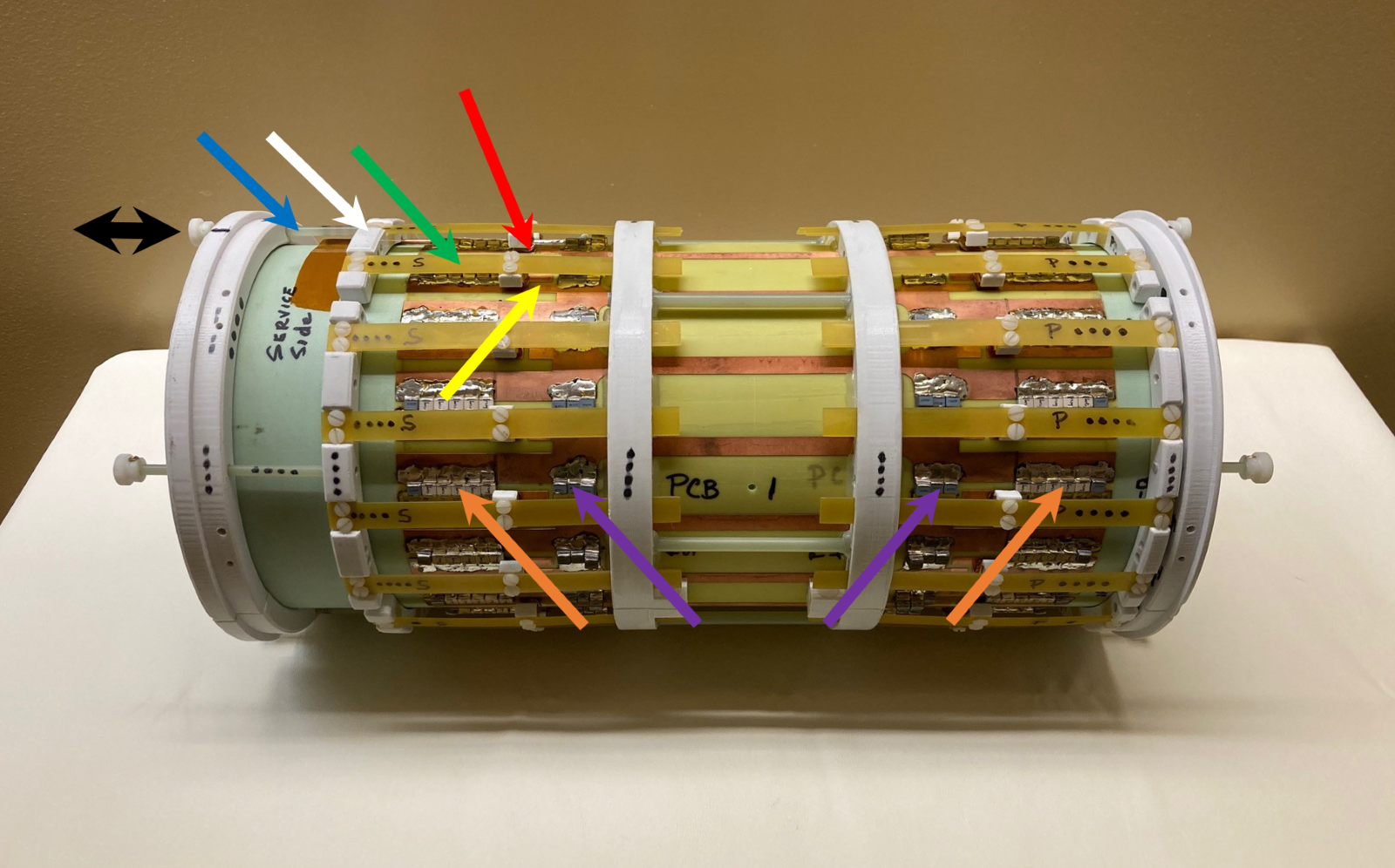
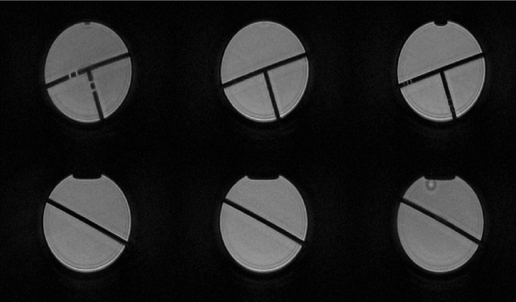
Figure 2. 1H 3D radial ultrashort echo time (UTE) axial images of a water filled cylindrical phantom.