3550
Robust spatial and temporal unwrapping for accurate quantitative susceptibility mapping
Alex Ensworth1, Véronique Fortier1,2, Jorge Campos Pazmino1, and Ives R Levesque1,2,3,4
1Medical Physics Unit, McGill University, Montreal, QC, Canada, 2Biomedical Engineering, McGill University, Montreal, QC, Canada, 3Research Institute of the McGill University Health Centre, Montreal, QC, Canada, 4Gerald Bronfman Department of Oncology, McGill University, Montreal, QC, Canada
1Medical Physics Unit, McGill University, Montreal, QC, Canada, 2Biomedical Engineering, McGill University, Montreal, QC, Canada, 3Research Institute of the McGill University Health Centre, Montreal, QC, Canada, 4Gerald Bronfman Department of Oncology, McGill University, Montreal, QC, Canada
Synopsis
Phase data for quantitative susceptibility mapping must be unwrapped to remove ambiguities due to limitations in the collected data. This work demonstrates the advantages of quality-guided region-growing over conventionally used methods and introduces a general method to correct for temporal discontinuities introduced by path-based spatial unwrapping methods. The unwrapping approaches were evaluated in 11 in vivo datasets presenting a variety of phase profiles.
Introduction
Accurate phase unwrapping is crucial for accurate quantitative susceptibility mapping (QSM). Path-following spatial unwrapping has been reported as more accurate than Laplacian approaches1. Despite this, studies continue to use Laplacian unwrapping for QSM2,3,4. This work studied the accuracy of phase unwrapping approaches for QSM, including a recently published spatial unwrapping technique, and temporal unwrapping. Our results show higher accuracy for path-following spatial unwrapping, but this approach can introduce temporal wraps, so a method is proposed to remove these.Methods
Six spatial unwrapping techniques were compared, and a temporal phase unwrapping method was developed (Temporal pHase Unwrapping by Median Phase Restoration, or THUMPR). Three Laplacian-based techniques (obtained from the morphology enabled dipole inversion (MEDI) toolbox5 and the susceptibility tensor imaging (STI) suite6, and one obtained from a collaborator) and three path-following techniques (quality guided region growing (QGRG)1, region growing from the MEDI toolbox, and Speedy rEgion-Growing Algorithm for Unwrapping Estimated Phase (SEGUE)7) were included. THUMPR was designed in response to ad hoc temporal wraps introduced when using path-following spatial unwrapping. THUMPR identifies large phase jumps between entire spatially unwrapped volumes at successive echoes by considering the progression of the median phase over time, and applying 2π corrections where necessary. The study was performed using 11 datasets acquired on two 3 T systems in four otherwise healthy volunteers (three males aged 26, 26, and 43, and one female aged 25) under institutional ethics approval. All data were acquired with vendor-provided multi-echo gradient echo pulse sequences using multi-channel head coils. Acquisition parameters were varied slightly for each dataset (Table 1). Susceptibility maps were produced using projection onto dipole fields (PDF)8 for background field removal and MEDI5 for dipole inversion. Spatial unwrapping abilities were compared quantitatively by line profile extraction and qualitatively on the final susceptibility map.Results
QGRG spatial unwrapping generally performed best; SEGUE also performed very well and was considerably faster than QGRG. Each implementation of Laplacian-based unwrapping generated different results, and all modified the original phase profile. These results can be seen in Fig. 1. Region growing failed in certain datasets and SEGUE failed in regions of rapidly varying phase. QGRG resulted in no spatial unwrapping failures in these 11 datasets. THUMPR detected and corrected temporal wraps present in this study, such as in the example in Fig. 2, consistently for all 11 datasets. Using the mean instead of the median as a metric for THUMPR was found to behave similarly. Poor spatial unwrapping and uncorrected temporal wraps corrupted the quantitative susceptibility maps as expected (Figs 3 and 4, respectively).Discussion
QGRG and SEGUE provided accurate spatial unwrapping that was consistent with the measured data, suggesting that path following methods are superior, except for isolated failures with SEGUE. Phase alterations resulting from Laplacian unwrapping can result in poor background removal and inaccurate susceptibility estimates. These results were consistent across 11 ad hoc datasets and are therefore likely to be generalized. Short echo spacing greatly reduces the likelihood of natural temporal phase wraps in the brain by reducing overall inter-echo phase accumulation. Path following spatial unwrapping can introduce apparent temporal phase wraps depending on the spatial location of the starting point for unwrapping: if two successive echoes are unwrapped from starting points in different locations, the initial position of spatial wraps can introduce 2π jumps between these echoes. THUMPR was able to robustly remove any such wraps, natural or apparent, in all datasets in this study. THUMPR relies on restoring the temporal progression of the median phase in spatially unwrapped data and does not impose constraints on echo spacing8.Conclusion
This work suggests that QGRG spatial unwrapping paired with THUMPR should be preferred over Laplacian unwrapping techniques for QSM, to ensure that the unwrapped phase data is accurate and consistent with the original wrapped phase.Acknowledgements
This research was funded by CIHR (Canadian Institutes of Health Research). The authors acknowledge Guillaume Gilbert (Philips healthcare) for his software on echo combination and Laplacian unwrapping as well as the Cornell MRI Research lab (MEDI toolbox and online software) and the Brain Imaging and Analysis Center from Duke University (STI suite)for access to their QSM toolboxes, and the MRI Research Lab at the University College London for access to SEGUE.References
1. V. Fortier and I. R. Levesque, “Phase Processing for Quantitative Susceptibility Mapping of Regions With Large Susceptibility and Lack of Signal,” Magn. Reson. Med., vol. 79, no. 6, pp. 3103–3113, 2018.2. S. Bollmann et al., “DeepQSM - using deep learning to solve the dipole inversion for quantitative susceptibility mapping,” Neuroimage, vol. 195, pp. 373–383, 2019.
3. J. Yoon et al., “Quantitative susceptibility mapping using deep neural network: QSMnet,” Neuroimage, vol. 179, no. 1, pp. 199–206, 2018.
4. M. W. A. Caan, et al., “MP2RAGEME: T1, T2*, and QSM mapping in one sequence at 7 tesla,” Hum. Brain Mapp., vol. 40, no. 6, pp. 1786–1798, Apr. 2019.
5. J. Liu et al., “Morphology enabled dipole inversion for quantitative susceptibility mapping using structural consistency between the magnitude image and the susceptibility map,” Neuroimage, vol. 59, no. 3, pp. 2560–2568, 2012.
6. W. Li, B. Wu, and C. Liu, “Quantitative susceptibility mapping of human brain reflects spatial variation in tissue composition,” Neuroimage, vol. 55, no. 4, pp. 1645–1656, 2011.
7. A. Karsa and K. Shmueli, “SEGUE: A Speedy rEgion-Growing Algorithm for Unwrap- ping Estimated Phase,” IEEE Trans. Med. Imaging, vol. 38, no. 6, pp. 1347–1357, 2019.
8. S. Robinson et al., “A method for unwrapping highly wrapped multi-echo phase images at very high field: UMPIRE,” Magn. Reson. Med., vol. 72, no. 1, pp. 80–92, 2014.
Figures
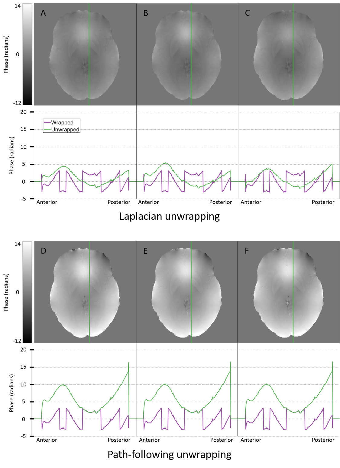
Six different techniques (three Laplacian, top row, and three path-following, bottom row) were used to spatially unwrap phase data from dataset C2. The output from each unwrapping technique is shown with an axial cross-section of the brain, and a line profile beneath it. Panel A represents the Laplacian from the STI suite, B is the Laplacian from the MEDI toolbox, C is the Laplacian from a collaborator, D represents QGRG, E represents SEGUE and F represents region growing from the MEDI toolbox.
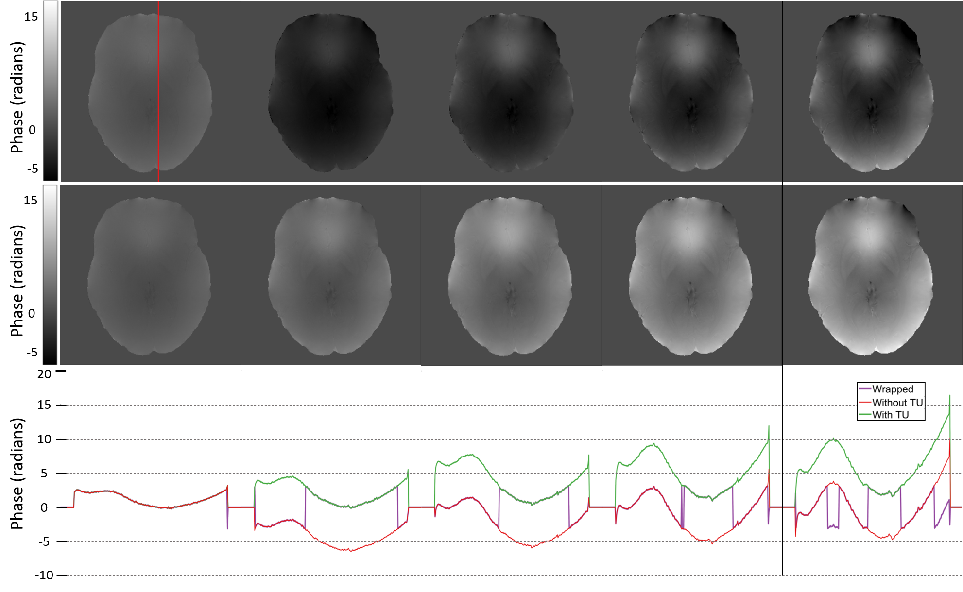
Five echoes after QGRG spatial unwrapping are shown across the columns. From left to right, the first echo is at 5.0 ms, followed by 10.4 ms, 15.8 ms, 21.2 ms and then 26.6 ms. The top row is without temporal unwrapping (TU), the middle row is with temporal unwrapping and the bottom row is a plot of the line profiles for each column. Dataset C2 was used.

Susceptibility maps which compare Laplacian and QGRG unwrapping techniques are shown. Laplacian unwrapping is the left column, QGRG unwrapping is in the middle, and their difference is shown in the rightmost column. Dipole inversion was done using MEDI, with data from dataset C2.
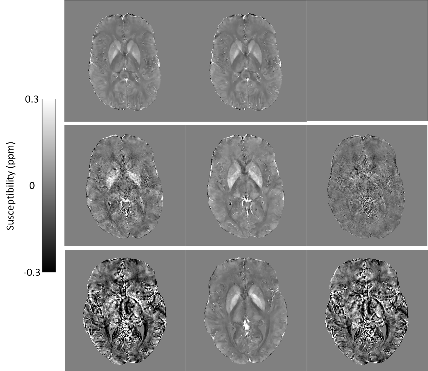
Susceptibility maps comparing the effects of temporal unwrapping across different datasets are shown. QGRG was used for spatial unwrapping and MEDI was used for dipole inversion. In the left column, no temporal unwrapping is used, in the middle column temporal unwrapping is used and the right most column is their difference. Each row is a different dataset, from top to bottom: B1, B2 and C2.
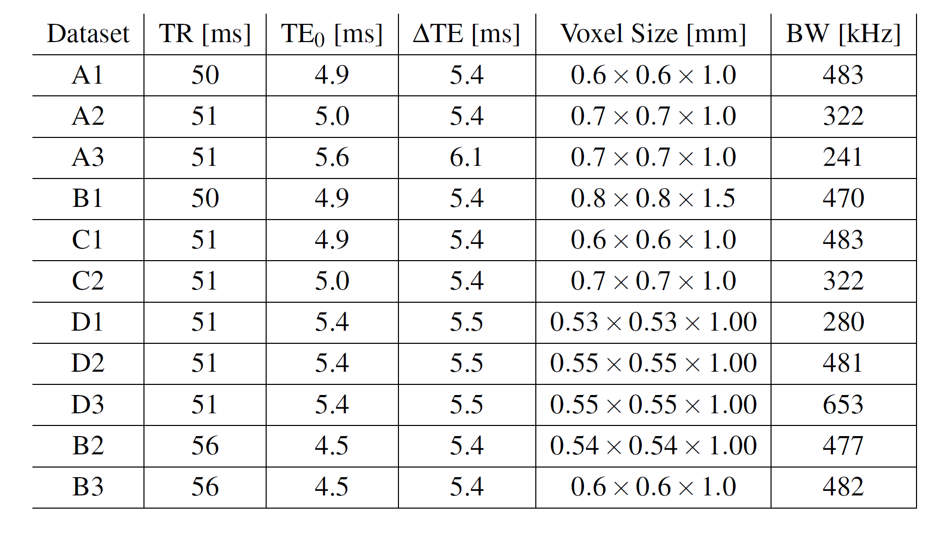
The scan parameters for in-vivo data acquisition across four volunteers. All datasets were acquired on a Philips system, except dataset B1 on a Siemens system. All datasets had 5 echoes, except dataset B3 which had 7. The letters correspond to volunteers and the numbers to specify the dataset.