3548
Evaluation of noise reduction performance using deep learning reconstruction: A phantom study1Fukushima Medical University, Fukushima, Japan, 2Hoshi General Hospital, Koriyama, Japan
Synopsis
We aim to assess fundamental noise reduction performance using deep learning reconstruction with a phantom at a 1.5 T MR scanner. In this study, the relationship among parameters for noise reduction, signal-to-noise ratio, image quality, and spatial resolution of images was evaluated. SNRs were increased higher significantly by DLR in all SNR ranges. Increasing ratio of SNR was varied by means of parameter settings. Combination of the DLR parameters affected varies to SNR, SSIM, and spatial resolution of the images. We should exercise caution to select DLR parameters when this technique applies to clinical images.
[Introduction]
Image quality in magnetic resonance (MR) is based on the MR physics, sometimes happens some conflicts of parameters to increase image quality such as signal-to-noise ratio (SNR), spatial resolution, scan time, and so on. (1) Strength of magnetic field is one of the parameters for defining SNR, it is very difficult to change it to increase SNR. (2) Deep learning is a branch of artificial intelligence and has been developing a lot of algorithms to solve complex problems related to medical images. Deep learning reconstruction (DLR) (Advanced Intelligent Clear-IQ Engine {AiCE}, Canon Medical Systems, Tokyo, Japan) was the first commercialized DLR tool to reduce noise of MR images. (3) It has some parameters to manage the strength of noise reduction, and we have little evidence to refer to determine parameters. The purpose of this study was to clarify fundamental characteristics of this tool using a phantom to provide properties for clinical use.[Methods]
1.5T MR equipment (Vantage Orian, Canon Medical Systems) with body and spine Coils was used. Type 90-401 phantoms containing polyvinyl alcohol (PVA) gel (Nikko Fines Industries Co. Ltd.) was used to evaluate image quality. 2-dimensional spin-echo (SE) sequence with TR = 500 ms., TE = 15 ms., number of average = 1, pixel bandwith = 217 Hz, FOV = 25.6 x 25.6 cm2, matrix = 368 x 368, spatial resolution = 0.7 x 0.7 mm2, scan time = 3:05. Images with 8 mm, 4 mm, and 2 mm of slice thickness were obtained to change SNR. 6 slices of axial images with the gap more than 2 times wider slice thickness were obtained for each phantom section. After obtained these images, DLR was applied to reduce noise with various parameters. To evaluate image quality, ROIs were set on the images and measure mean and SD on them. Although some SNR calculation methods have been proposed in the past literature, SNR was defined as a mean value divide by SD on the same ROI in this study. (4-6) SNRs were calculated on each ROI on each slice with various DLR parameters. Structural similarity (SSIM) were calculated using MATLAB R2020b. In addition to these analyses, visual evaluation with a scoring method of pin section was performed to evaluate changes of spatial resolution by four radiological technologists who have various experience of clinical MR exam. All statistical analyses were performed with SPSS statistics 27. Repeated-measures-ANOVA were performed to detect significant differences. Then Friedman test was performed as a posthoc test.[Results]
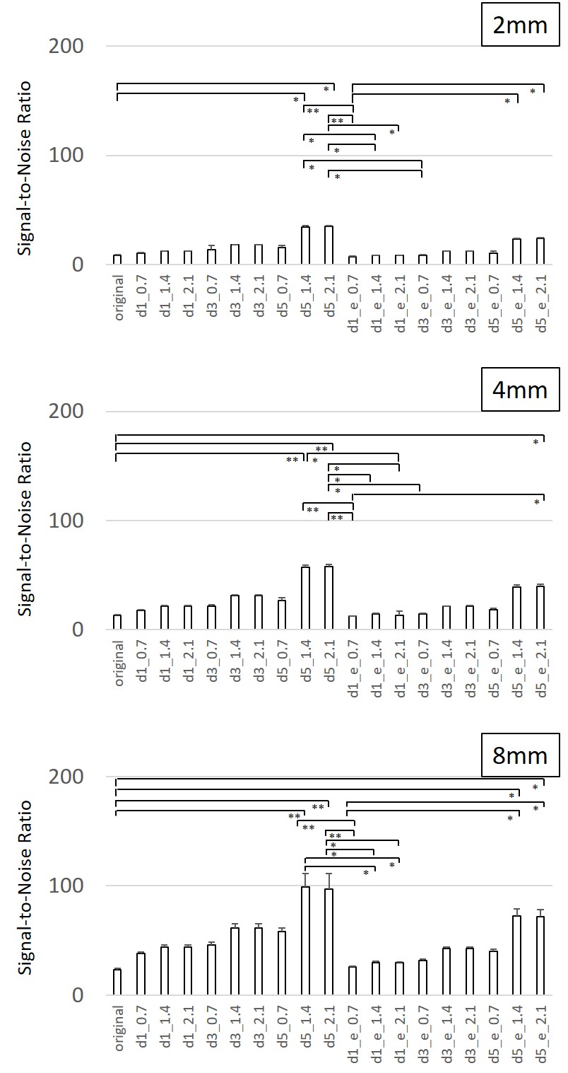
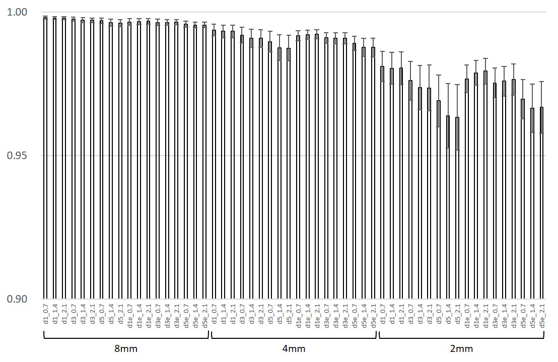
Figure 1 shows SNR changes in terms of DLR on images with 2 mm, 4 mm, and 8 mm slice thickness, respectively. Original indicated without DLR. DLR made SNR of images higher in comparison to that of original images. Figure 2 shows SSIM changes in terms of DLR. Decreasing of SSIM was shown in more thin slices. Edge enhancement function recovered SSIM higher, especially in the lower SSIM parameters. The results of visual estimation showed that mean scores were recovered by using edge enhancement, especially in the phase encoding direction compared to that in the readout direction.[Discussion]
The fundamental noise reduction performance of DLR was evaluated using a phantom in this study. SNRs were increased higher significantly by DLR in all SNR ranges. Increasing ratio of SNR was varied by means of parameter settings. Decreasing of SSIM higher in lower SNR images is compared to that in higher SNR images. Edge enhancement recovered SSIM higher, especially in lower SNR images. The results of visual estimation indicated that little changes of a mean score by means of parameters were shown and edge enhancement has little effects on improvement of spatial resolution. Different trends of mean score of visual evaluation by means of the DLR parameters between phase encode direction and readout direction of the images were found.[Conclusion]
DLR had a powerful performance to increase SNR in all SNR ranges. Combination of the DLR parameters affected varies to SNR, SSIM, and spatial resolution of the images. We should exercise caution to select DLR parameters when this technique applies to clinical images.Acknowledgements
No acknowledgement found.References
1. Saloner D, et al., Magn Reson Imaging Clin N Am. 2015;23(1):1–6.
2. Rutt BK, et al., JMRI. 1996;1:57-62.
3. Kidoh M, et al., Magn Reson Med Sci. 2020;19(3):195-206.
4. NEMA Standards Publication MS 1-2008 (R2014)
5. Dietrich O, et al., J Magn Reson Imaging. 2007;26:375-385.
6. Goerner FL, et al., Med Phys. 2011;38(9):5049-5057
Figures