3547
Usefulness of Deep Learning Based Denoising Method for Compressed Sensing in Pituitary MRI
Takeshi Nakaura1, Hiroyuki Uetani1, Kousuke Morita1, Kentaro Haraoka2, Akira Sasao1, Masahiro Hatemura1, and Toshinori Hirai1
1Diagnostic Radiology, Kumamoto University, Kumamoto, Japan, 2Cannon Medical Systems Japan, Tochigi, Japan
1Diagnostic Radiology, Kumamoto University, Kumamoto, Japan, 2Cannon Medical Systems Japan, Tochigi, Japan
Synopsis
We evaluated image quality of hybrid type deep learning reconstruction (hybrid-DLR) with wavelet based denoising method in T2-weighted images (T2WI) of the pituitary with various denoising level (1-5). There was a progressive increase in SNR with hybrid-DLR with increase of the denoising level. On the other hand, the SNR of conventional wavelet-based method was not increased at high denoising levels (4-5). All qualitative scores of hybrid-DLR in any denoising levels are higher than that of wavelet based denoising method, and the difference became more noticeable at higher denoising levels.
Introduction
Compressed sensing (CS) in MRI has been reported that it is possible to reconstruct images from fewer measurements than required in traditional sampling method if some constraints are satisfied. In this technique, an iterative reconstruction (IR) with a wavelet filter has been used to improve the SNR. However, previous reports have suggested that this technique has the possibility to shows global ringing artifacts and blurring of fine details at high denoising level 1,2. Recently, the usefulness of deep learning-based reconstruction (DLR) for denoising MRI images has been reported 3,4. DLR denoising is a technology based on convolutional neural networks applied to image denoising and can be used for denoising reconstructed images by IR with wavelet filter. Therefore, we hypothesized that DLR based denoising method might be useful to denoise the noisy images reconstructed by IR with wavelet filter from fewer measurements. The purpose of this study was to evaluate the efficacy of hybrid type DLR for the pituitary MRI.Methods
This retrospective study included 19 consecutive patients who underwent T2 weighted images (T2WI) using a 3T MRI scanner (Vantage Galan 3T ZGO; Canon Medical Systems, Kanagawa, Japan) with a 32-channel head coil. The parameters of T2WI were as follows: Repetition time (ms), 4000; Echo time (ms), 93.5; Echo train lengths 21; Echo space (ms) 8.5; Flip angle 90/160°; Matrix 224×272; Field of view 130×130 mm; Slice thickness/gap (mm) 3/0.3; No. of slices 11; Bandwidth (Hz) 325.5; Acceleration factor 2.4; NAQ 2.0; Acquisition time (s) 1:08. The images were reconstructed with the conventional wavelet based denoising method or the DLR based denoising method at various denoising levels (1-5). Denoising levels were changed in the conventional wavelet based denoising method using the regularization factor (1.4, 1.7, 2.0, 2.4 and 2.7), and were changed in the DLR based denoising method using the blending rate (30%, 40%, 50%, 60% and 70%). The SNRs of the cerebrospinal fluid (CSF), callosum and pons, contrast between the CSF and pons, the CSF and callosum, and the callosum and pons were compared between the two image types. Image noise, sharpness, artifacts, and overall image quality of these two types of images were scored using a 4-point grading scale.Results
Figure 1 shows results of SNR and Contrast with the conventional wavelet based denoising method or the DLR based denoising method at various denoising levels. There was a progressive increase in SNR of CSF with hybrid-DLR with increase of the denoising level for CSF (Level 1-5: 27.18±8.42, 30.03±9.99, 33.54±12.37, 38.28±15.79 and 43.17±19.48). On the other hand, the SNR of CSF with conventional wavelet-based method was not increased at high denoising levels (Level 1-5: 26.14±7.89, 28.87±9.41, 30.88±10.66, 30.08±10.04 and 26.82±7.66). Similar results were observed for SNR of callosum and pons. The differences of the contrast between the two denoising approaches were small. Figure 2 shows results of qualitative analysis with the conventional wavelet based denoising method or the DLR based denoising method at various denoising levels. In qualitative analyses, all qualitative scores of hybrid-DLR in any denoising levels are higher than that of wavelet based denoising method. The difference of qualitative scores between the two denoising approaches became more noticeable at higher denoising levels.Conclusion
In conclusion, the DLR based denoising method can improve the image quality of T2WI of pituitary with CS as compared with the conventional wavelet based denoising method. The difference between the two denoising approaches became more noticeable at higher denoising levels.Acknowledgements
None.References
1. Sharma SD, Fong CL, Tzung BS, Law M, Nayak KS. Clinical image quality assessment of accelerated magnetic resonance neuroimaging using compressed sensing. Investigative radiology 2013;48(9):638-645.2. Zhang T, Chowdhury S, Lustig M, et al. Clinical performance of contrast enhanced abdominal pediatric MRI with fast combined parallel imaging compressed sensing reconstruction. J Magn Reson Imaging 2014;40(1):13-25.
3. Jiang D, Dou W, Vosters L, Xu X, Sun Y, Tan T. Denoising of 3D magnetic resonance images with multi-channel residual learning of convolutional neural network. Jpn J Radiol 2018;36(9):566-574.
4. Zhang K, Zuo W, Chen Y, Meng D, Zhang L. Beyond a Gaussian Denoiser: Residual Learning of Deep CNN for Image Denoising. IEEE transactions on image processing : a publication of the IEEE Signal Processing Society 2017;26(7):3142-3155.
Figures
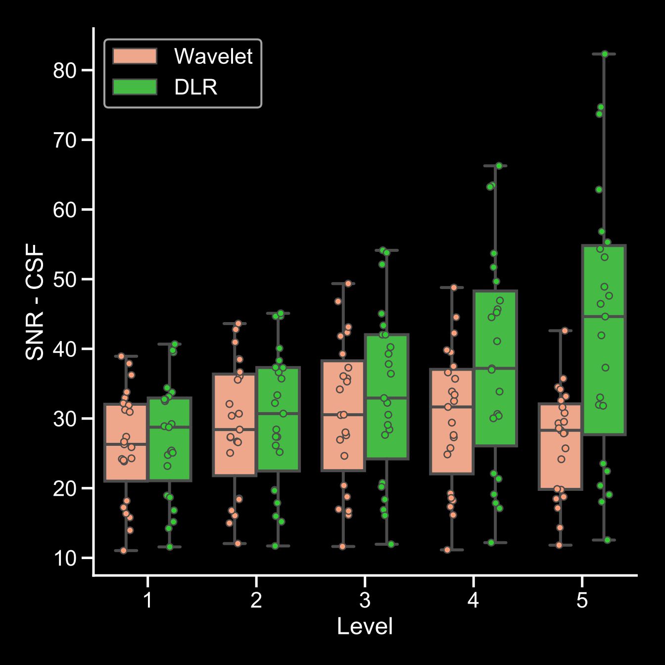
A box plot shows the SNR of CSF. There was a progressive increase in SNR of CSF with DLR
based denoising method with increase of the
denoising level in any location. On the other hand, the SNR of conventional
wavelet-based method was not increased at high denoising levels (4-5).
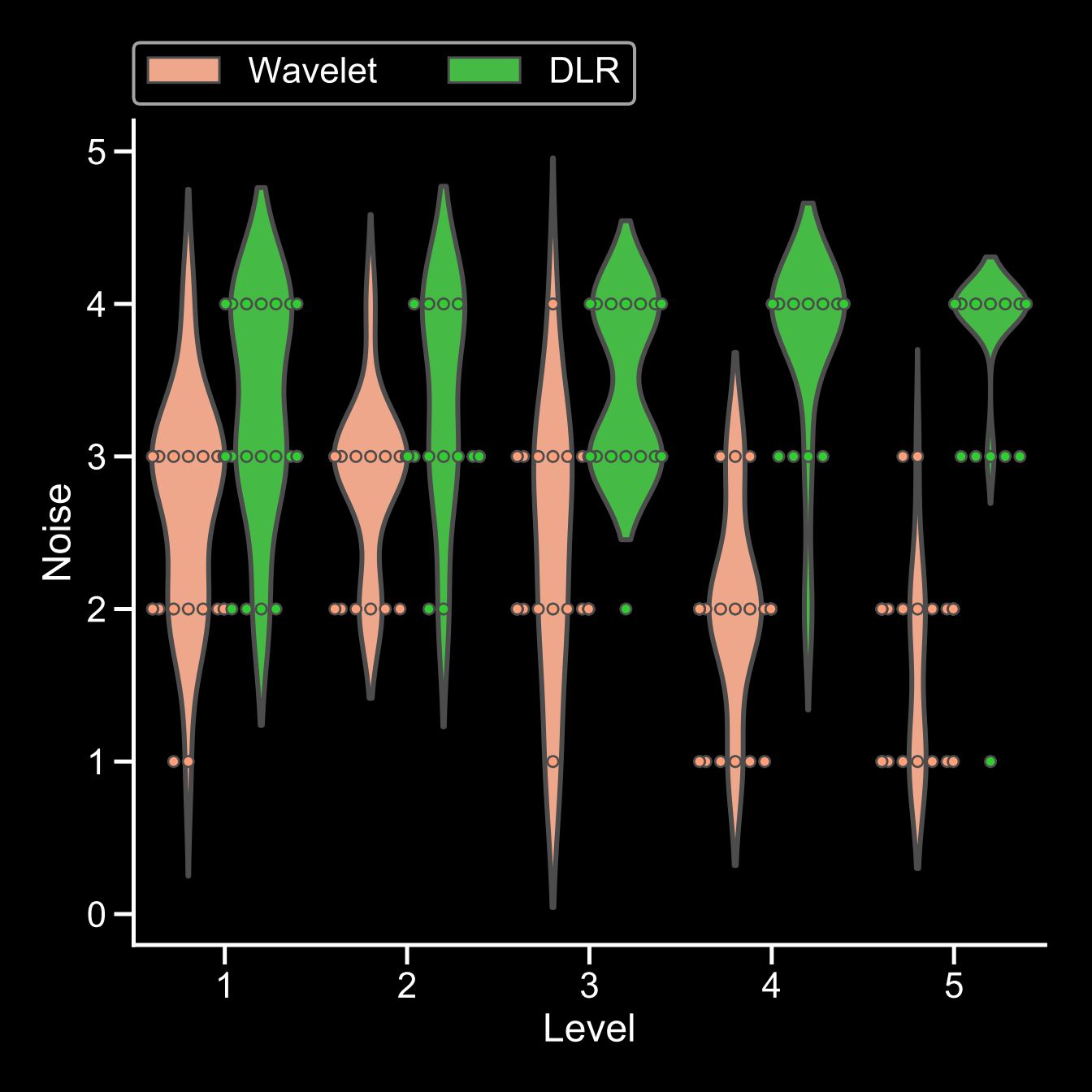
A violin plot shows the result about the over-all image quality. All
qualitative scores of DLR based denoising method in
any denoising levels are higher than that of wavelet based denoising method,
and the difference became more noticeable at higher denoising levels.
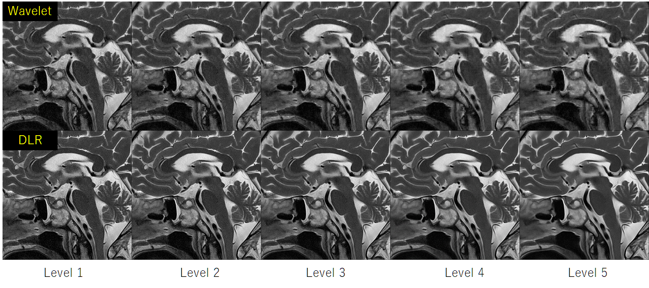
Representative case. (upper row) Conventional wavelet based denoising
method, (lower row) DLR based denoising
method. The DLR based denoising method offered decrease in image noise,
artifact, and clear depiction of pituitary grand and small objects as compared
with the conventional wavelet based denoising method.