3543
Image blending method to produce consistent SNR in images denoised using a convolutional neural network1Magnetic Resonance, Canon Medical Research USA, Inc., Mayfield Village, OH, United States
Synopsis
Deep learning based denoising methods can significantly reduce image noise at the cost of producing unnaturally smooth images. Typically, natural-looking images are produced by blending a fraction of the acquired noisy image with the denoised image. The blending ratio is set based on visual inspection. This approach impedes workflow and is prone to inter-operator variability. We propose a method to analytically calculate the blending ratio based on a desired SNR value. The proposed method is demonstrated to produce natural-looking denoised images with consistent SNR across head, spine and knee applications.
Introduction
In recent years, several deep learning-based methods have been proposed to denoise high resolution MR images1-3. Typically, a convolutional neural network (CNN) is used. The CNN is trained on a range of noise levels to allow denoising of a wide variety of clinical images containing different amounts of noise. A common challenge in these deep learning methods is that the denoised images appear unnaturally smooth. Even when all the anatomical details and image contrast are preserved, the denoised image can look artificially plastic. Clinicians trained to read medical images tend to prefer a natural-looking image, which includes some amount of image noise. The complete absence of noise can impede diagnostic interpretation. To provide a natural looking image, the denoised image is typically blended with a fraction of the original noisy image. The blending ratio is empirically set by the operator based on visual inspection. This qualitative approach impedes workflow and is vulnerable to inter-operator variability.We propose a method to automatically blend the output denoised images with the input noisy images to generate images with consistent SNR across different anatomies and contrasts. Instead of qualitatively setting the blending ratio for each imaging volume, a common target SNR value can be set for a range of scan conditions. The target SNR value is used to analytically calculate the blending ratio without operator intervention. We demonstrate that the proposed method can produce consistent SNR in images denoised using a CNN across head, spine, and knee applications.
Methods
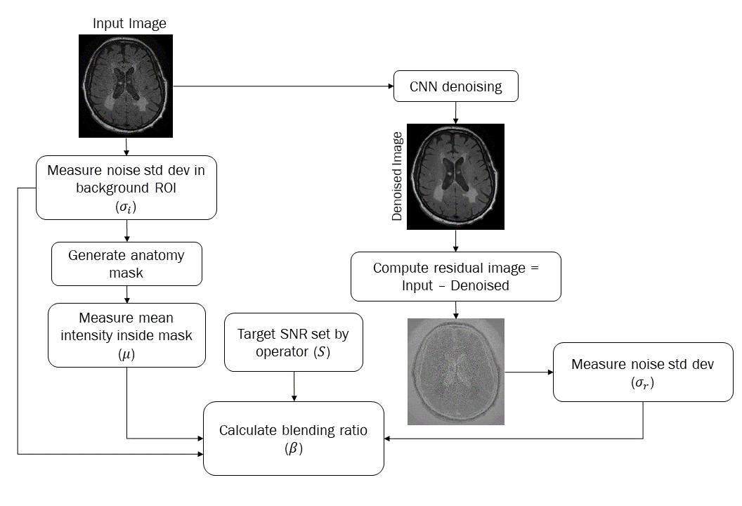
The blending ratio (β) is calculated using the formula $$$\beta = 1 - \frac{S\sigma_i-\mu}{S\sigma_r}$$$ where S is the target SNR value, μ is the mean image intensity in the tissue voxels, σi is the noise standard deviation in the input noisy image, σr is the noise standard deviation in the residual image. Figure 1 shows the steps used to calculate β. For a given noisy input image Inoisy and denoised image Idenoised, the blended image is given by Iblended = Idenoised + βInoisy.To test the proposed method, a residual CNN motivated by DnCNN4 was designed with 19 hidden and 2 outer layers. The input layer comprised a convolution layer followed by ReLU activation. The 19 hidden layers comprised a convolution layer with ReLU activation and batch normalization, followed by an output convolution layer. All layers had 64 convolution kernels of size 3x3. A residual connection was made between the input and output layers.
Training images were acquired in normal subjects on a Vantage Galan 3T scanner (Canon Medical Systems Corporation, Japan) using head T1-weighted FSE, T2-weighted FSE, FLAIR, and MP-RAGE sequences. Seven studies were acquired using each sequence, for a total of 28 studies. Typical FOV was 24x24 cm2. Acquisition matrix size was varied between 256x256 and 640x640 to obtain training images at different spatial resolutions. Training image pairs of low SNR input and high SNR target images were created. The high SNR target images were obtained by rigid-body registration and averaging of five repeated measurements of the same imaging volume. The low SNR input images were generated by adding noise to high SNR target images. Noise with levels in the range 3-25% was added based on measurements performed on clinical protocols. Noise level is defined by the noise standard deviation as a percentage of the mean image intensity inside the anatomy of interest.
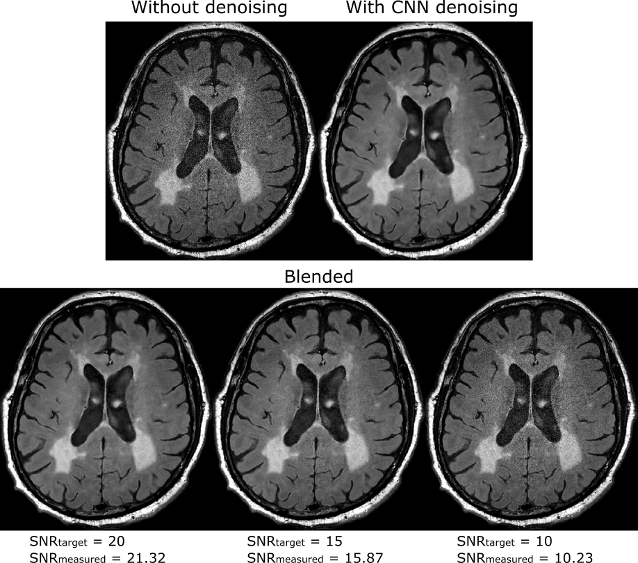
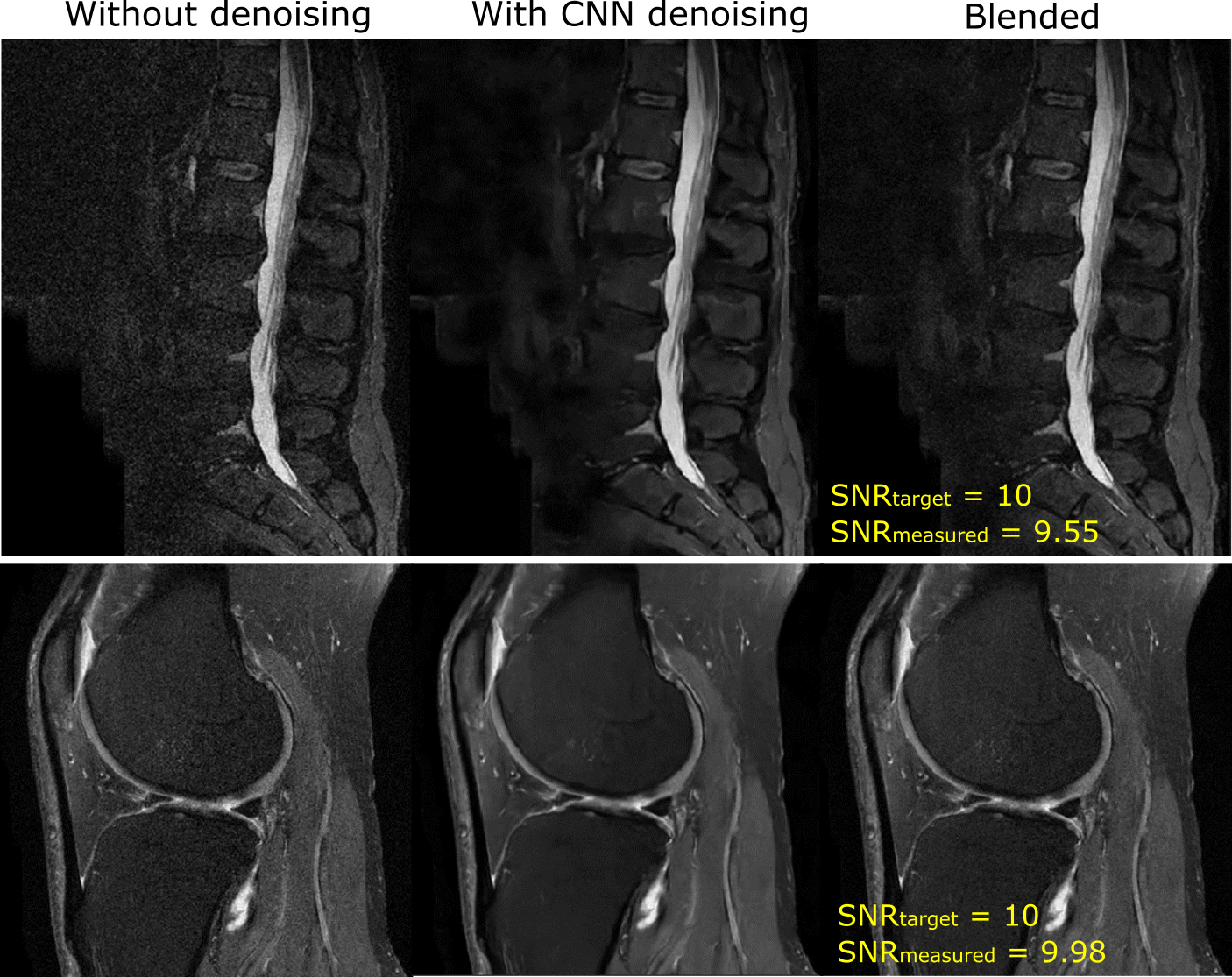
Test images were acquired using head axial FLAIR, lumbar spine sagittal STIR, and fat suppressed knee sagittal intermediate-weighted (IW) sequences. The test images were denoised using the trained CNN. The denoised images were blended with the input images using the proposed method. The head images were blended with target SNR values of 10, 15 and 20. The knee and spine images were blended with a target SNR value of 10.
Results
Figure 2 shows the results from the head FLAIR experiments. Images processed by the denoising CNN have high SNR and contain all the anatomical details but appear unnaturally smooth. The blended images demonstrate the application of the proposed method at different target SNR values. The SNR measured on the blended images closely match the target SNR values. Figure 3 shows the results from lumbar spine STIR and knee IW experiments. The images denoised by the CNN have high SNR compared to images without denoising. However, the images produced by the CNN appear undesirably smooth. The blended images produced by the proposed method provide a good balance between SNR improvement and image smoothness. The SNR values measured on the blended images closely match the target SNR values.Discussion
We have demonstrated a method that provides consistent SNR in images denoised using a convolutional neural network, across head, lumbar spine, and knee applications. Instead of using visual inspection to set the blending ratio, the proposed method utilized SNR as a quantitative metric to produce images of consistent diagnostic quality across different applications. The benefits of the proposed method can be further verified in a future clinical validation study.Acknowledgements
No acknowledgement found.References
1. Kidoh M, Shinoda K, Kitajima M, et al. Deep learning based noise reduction for brain MR imaging: tests on phantoms and healthy volunteers. Magn. Reson. Med. Sci. doi:10.2463/mrms.mp.2019-0018.
2. Zhao Y, Liu Y, Mak HK, et al. Simultaneous denoising of multi-contrast MR images using a novel weighted nuclear norm minimization approach. ISMRM 27th annual meeting 2019, p. 0669.
3. Jiang D, Dou W, Vosters L, et al. Denoising of 3D magnetic resonance images with multi-channel residual learning of convolutional neural network, arXiv:1712.08726v2, 2018. Zhang K, Zuo W, Chen Y, et al. Beyond a Gaussian denoiser: residual learning of deep CNN for image denoising. arXiv:1608.03981v1, 2016.
Figures