3530
Simultaneous super resolution and distortion correction for Single-shot EPI DWI using deep learning method1Center for Biomedical Imaging Research, Department of Biomedical Engineering, School of Medicine, Tsinghua University, Beijing, China, 2MR Clinical Science, Philips Healthcare, Suzhou, China
Synopsis
Single-shot EPI is widely used for clinical DWI acquisitions. However, due to the limited bandwidth along PE direction, the obtained images suffer from distortion and blurring, which limits its diagnosis capability. Here we propose a deep learning-based method to simultaneously increase resolution and correct distortions for SSh-EPI. In-vivo DWI data are used to test the proposed method. The results show that distortion-corrected high-resolution DWI images can be reconstructed from low-resolution SSh-EPI images and high-resolution anatomical images.
Introduction
Single-shot EPI is a commonly used sequence for diffusion-weighted MRI. Nevertheless, single-shot EPI suffers from geometric distortion and T2* blurring which limits the resolution. Many efforts have been made to tackle these problems1-3. Among them, PSF-EPI method can acquire distortion- and blurring-free high-resolution DWI images. However, these techniques may increase scan time. Deep learning-based methods have recently been proposed to transfer information from high-quality images to low-quality images without adding scan time4-6. Here we propose a deep learning network to transfer information from high-resolution distortion-free PSF-EPI DWI and anatomical T2W images to low-resolution SSh-EPI DWI images.The method can simultaneously increase resolution and correct distortions for SSh-EPI.Theory
EPI Geometric deformationPSF-EPI uses an additional phase encoding to obtain distortion-free images. For PSF-EPI acquisition, the signal acquisition expression can be written as:
$$S(k_y,k_s)=\int \rho (r)e^{-\frac{t_y}{T_2^*}}e^{ik_yr}e^{i\int_{0}^{t_y}\omega(r,t)dt}e^{ik_sr}dr (1)$$
Where $$$t_y=\frac{ESP*k_y}{\triangle ky}$$$, $$$k_s$$$ denotes extra phase encoding dimension, ESP represents echo spacing, $$$\omega $$$ refers to off-resonance caused by field inhomogeneity.
The inverse Fourier of (1) gets:
$$I(y,s)=\rho (s)H(y,s) (2)$$
After an integration along y and s respectively, we can get the following expression:
$$I(y)=\int I(s)\frac{H(y,s)}{\int{H(y,s)}dy }ds (3)$$
Thus, the distorted image $$$I(y)$$$ equals the convolution of a PSF function with the distortion-free image $$$I(s)$$$, which allows us to use a CNN network to correct the distorted images.
Super resolution problem
Deep learning methods like CNN networks are widely used for SR, since they can better learn the nonlinear mapping function between low-resolution image $$$I_{LR}$$$ and its corresponding high-resolution image $$$I_{HR}$$$ than traditional learning-based SR methods. Thus we propose a multi-task 2D CNN network to simultaneously increase resolution and correct distortions for low-resolution SSh-EPI.
Network structure
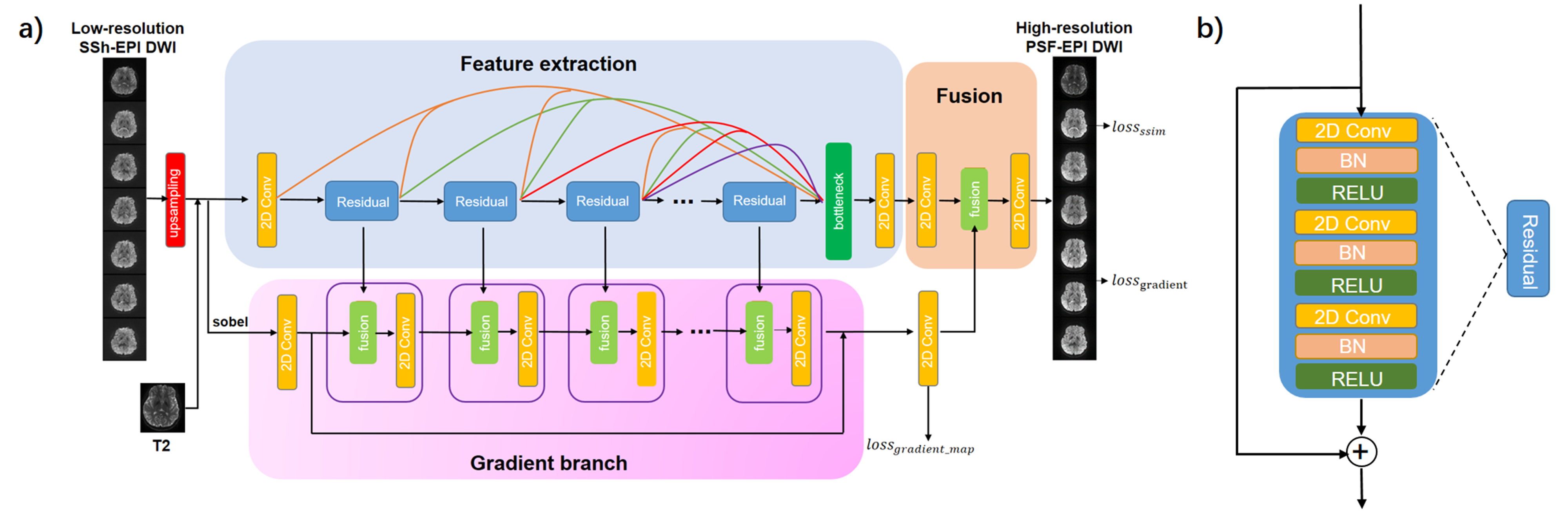
The network structure is shown in Fig. 1a. The feature extraction branch uses a densely connected 2D CNN network with multi-level fusion. Each input patch consists of 8 channels (including 1 b = 0 s/mm2, 6 b = 1000s/mm2 and 1 T2W-TSE). The structure of Residual block is shown in Fig. 1b. All information from different blocks are fused together. The gradient branch has gradient blocks composed of fusion and 2D Conv layers. The output of the gradient branch $$$I_{gradmap}$$$ is compared to the gradient map of HR PSF-EPI images to serve as an additional loss function . The fusion branch combines the multi-level feature maps with gradient maps to reconstruct images. The network tries to optimize the following loss:
$$\min_{\theta}l_{ssim}+\alpha_1\parallel\triangledown{I_{SR}}-\triangledown{I_{HR}}\parallel_1+\alpha_2\parallel{I_{gradmap}}-\triangledown{I_{HR}}\parallel_1$$
Where $$$\theta$$$ represents trainable parameters and $$$\alpha_i$$$ represents tradeoff parameters.
Method
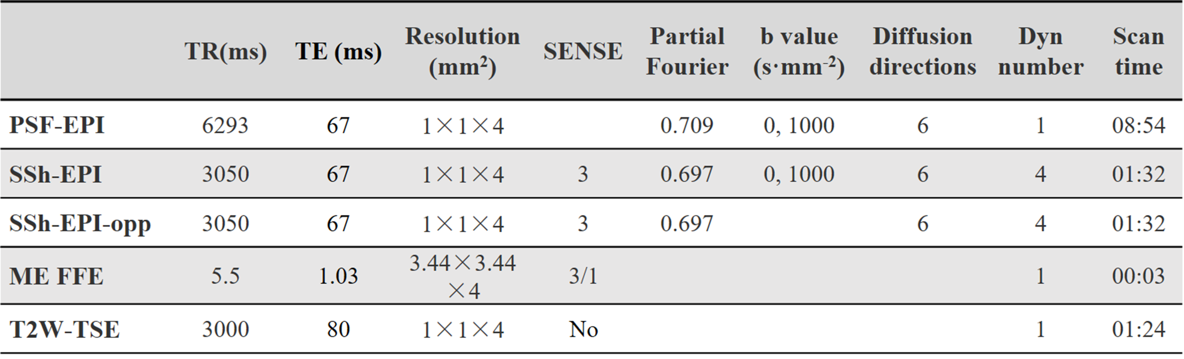
DatasetsIn vivo dataset: The data acquisition protocol can be found in Table 1. PSF-EPI DWI and T2W-TSE images were acquired with 1*1 mm2 in-plane resolution. SSh-EPI DWI images were downsampled to 2*2 mm2 in plane as input. The distortion levels remained consistent between interpolated LR SSh-EPI and HR SSh-EPI images. Multi-echo FFE was acquired to calculate field map and SS-EPI with opposite PE direction was acquired for the top-up correction. Twelve healthy volunteers were scanned on a Philips Ingenia 3T scanner (Philips Healthcare, Best, The Netherlands). We split subjects into 9, 2, 1 for training, validation and testing respectively. Each subject had 25 slices. The input LR patch size was 112*16 with 8 channels. The total number of paired patches for the training was 25000.
Evaluation
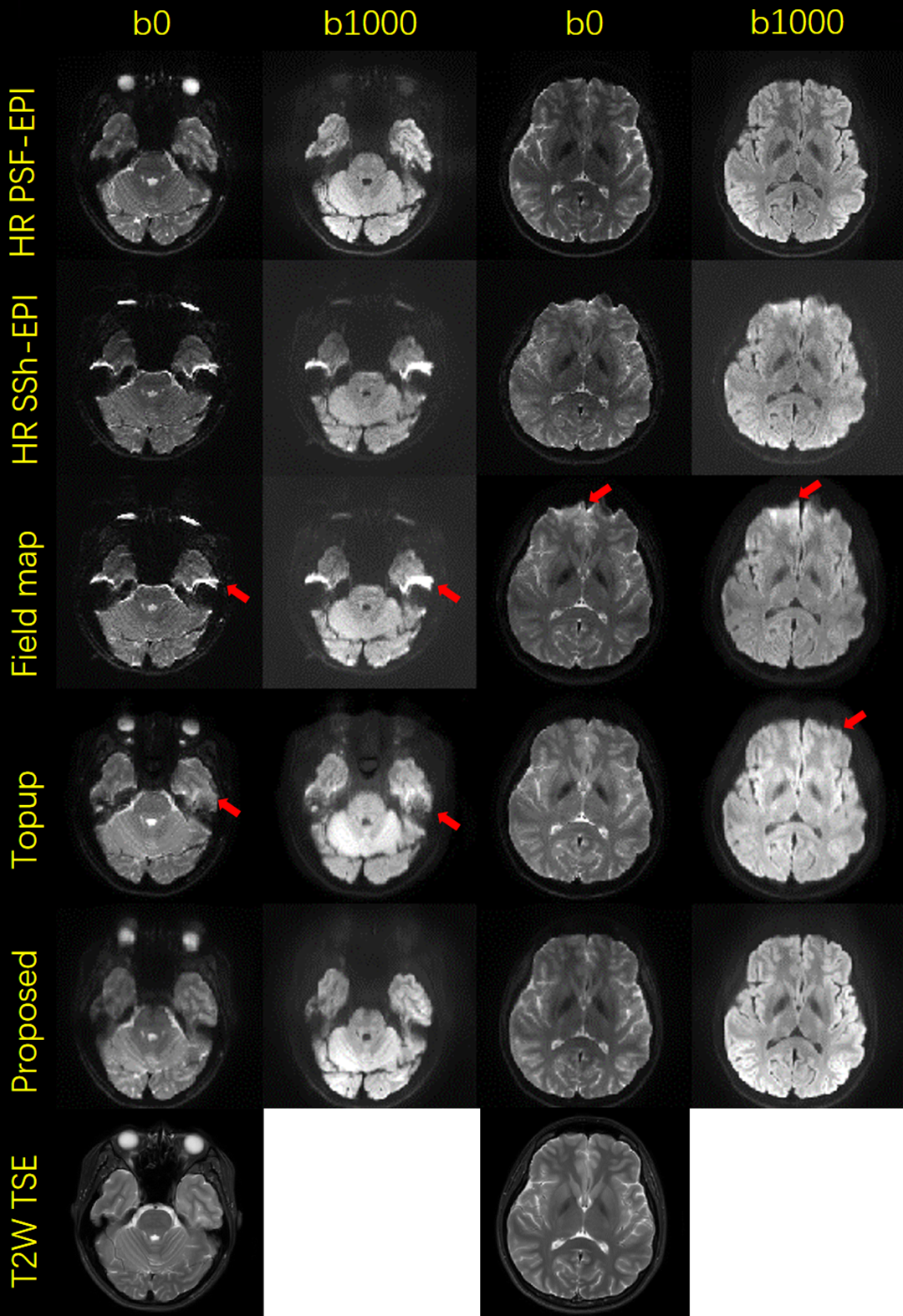
PSF-EPI data were used as reference. We used state-of-the-art field-mapping7 and top-up8 methods to correct distortions for original high-resolution SSh-EPI data. Then we compared the images reconstructed from low-resolution SSh-EPI using the proposed method to the results of field-mapping and top-up methods.
Results and Discussion
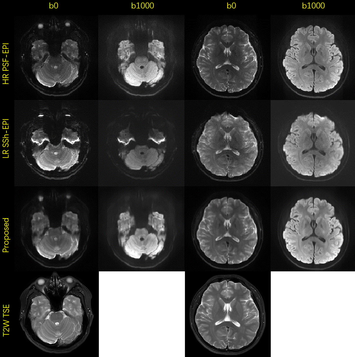
Figure 2 shows the distortion-corrected high-resolution b0 and mean DWI images obtained using the proposed method and the input low-resolution SSh-EPI images of one validation subject. Compared to input images, reconstructed images show sharper edges and the structures in regions suffering from severe field inhomogeneity can be restored. The results show that the proposed method can simultaneously increase resolution and correct distortions.Figure 3 shows the comparison results between different methods of the test subject. 2 representative slices of b0 and mean DWI images are selected. Though our proposed method uses low-resolution SSh-EPI as input, the results still show better consistency with the reference PSF-EPI. As shown in the images, strong signal pile-up still exists after field-mapping correction as pointed by the red arrowheads. The top-up method performs better than field-mapping, yet the fine structures cannot be restored well. Meanwhile, the proposed method could correct distortions and preserve detailed structure information.
Conclusion
We propose a deep learning method to simultaneously increase resolution and correct distortions for SSh-EPI. We demonstrate that the proposed method can reconstruct high-resolution distortion-corrected DWI images from low-resolution SSh-EPI images. Based on the results, the proposed method could better restore detailed structures consistent with the reference PSF-EPI.Acknowledgements
No acknowledgement found.References
1.Chen NK, Guidon A, Chang HC, Song AW. A robust multi-shot scan strategy for high-resolution diffusion weighted MRI enabled by multiplexed sensitivity-encoding (MUSE). Neuroimage 2013;72:41-47.
2.Mani M, Jacob M, Kelley D, Magnotta V. Multi-shot sensitivity-encoded diffusion data recovery using structured low-rank matrix completion (MUSSELS). Magnetic Resonance in Medicine 2017;78(2):494-507.
3.Dong ZJ, Wang FYX, Reese TG, et al. Tilted-CAIPI for highly accelerated distortion-free EPI with point spread function (PSF) encoding. Magnetic Resonance in Medicine 2019;81(1):377-392.
4.Ma C, Rao Y, Cheng Y, et al. Structure-Preserving Super Resolution with Gradient Guidance. Proceedings of the IEEE/CVF Conference on Computer Vision and Pattern Recognition 2020; 7769-7778.
5.Chen YH, Shi F, Christodoulou AG, Xie YB, Zhou ZW, Li DB. Efficient and Accurate MRI Super-Resolution Using a Generative Adversarial Network and 3D Multi-level Densely Connected Network. Lect Notes Comput Sc 2018;11070:91-99.
6.Hu Z, Wang Y, Zhang Z, et al. Distortion correction of single-shot EPI enabled by deep-learning. Neuroimage 2020;221:117170.
7.Jezzard P, Balaban RS. Correction for geometric distortion in echo planar images from B0 field variations. Magn Reson Med 1995;34(1):65-73.
8.Andersson JLR, Skare S, Ashburner J. How to correct susceptibility distortions in spin-echo echo-planar images: application to diffusion tensor imaging. Neuroimage 2003;20(2):870-888.
Figures