3521
Automated Radial Streaking Artifact Suppression with RGB-STAR1Medical Imaging, University of Arizona, Tucson, AZ, United States, 2Biomedical Engineering, University of Arizona, Tucson, AZ, United States, 3Electrical and Computer Engineering, University of Arizona, Tucson, AZ, United States
Synopsis
Streaking artifacts in radial MR imaging due to gradient nonlinearities are suppressed using a beamforming algorithm where region growing image segmentation is used to automatically generate the interference covariance matrix. The performance of the automatic streaking artifact suppression algorithm (RGB-STAR) is compared to algorithms based on coil removal and coil weighting and a beamforming algorithm with manual segmentation.
Introduction
Streaking artifacts that spread over the field-of-view (FOV) are a drawback of radial MRI techniques.1 Streaking artifacts are most commonly associated with azimuthal undersampling and motion but also arise as a consequence of gradient nonlinearities. This type of streaking artifact tends to originate from the anatomy at the periphery of the FOV, due to gradient imperfections close to the edge of the magnet. Since the level of artifact varies across the phased array RF coil elements,1 several coil selection and combination methods as well as phased-array processing algorithms have been proposed.2-5 Among these, the recently proposed beamforming for streaking artifact reduction (B-STAR) algorithm6 has the advantage that coils do not need to be removed or weighted, thus preserving the signal gains of phased array RF technology.6B-STAR estimates an interference covariance matrix using a region-of-interest (ROI) containing the artifact and then generates a phased array coil combination map using the signal correlation matrix at the spatial location and the fixed interference covariance matrix. B-STAR relies on the manual placement of the ROI around areas from which streaks emanate which is not practical for routine use, in particular if several ROIs need to be selected. The automatic generation of the interference covariance matrix by identifying the sources of streaking artifact can save time and produce more robust artifact reduction. Here, we present the RGB-STAR method which incorporates region growing image segmentation to automatically generate the interference covariance matrix which is then used to suppress streaking artifacts without reducing the signal in the anatomy of interest.
Methods
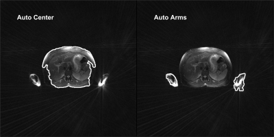
For the automatic selection of the ROIs, we used a region growing (RG) method7 with four seed points automatically placed at the four corners of the FOV of the intensity normalized image. RG selects the background area of the FOV (i.e., areas not including anatomical features). The complement of the segmented background region in abdominal MRI typically produces three foreground (or anatomical) regions based on their spatial location within the image: (i) the abdomen (region closest to the center of the image), (ii) the left arm (region closest to the bottom right of the image), and (iii) the right arm (region closest to the bottom left of the image) (Fig. 1). These foreground regions are used to generate the interference covariance matrix in two ways: a) the pixels in the complement of the central abdomen region (Auto Center) form the interference covariance matrix and b) the pixels in the union of the two arm regions (Auto Arms) form the interference covariance matrix.RGB-STAR was tested on a set of abdominal images acquired with a T2-weighted radial turbo spin echo pulse sequence. We selected 26 slices (from 13 subjects) with strong streaking artifacts originating from unsuppressed fat signal in the arms. The performance of RGB-STAR using the Auto Center and Auto Arm ROI selection was compared qualitatively and quantitatively against reference images (reconstructed with adaptive coil combination3 and without streak suppression) as well as images processed with a coil removal with outlier pruning (CR+OP) method4 and a sensitivity weighted (SW) coil combination method.5
Results
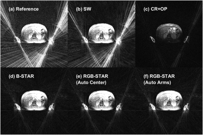
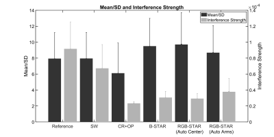
Figure 2 compares the level of streak removal achieved by RGB-STAR to the reference image and images processed with SW, CR+OP, and B-STAR. Note that the image reconstructed with SW has a similar level of artifact as the reference image. CR+OP suppressed the streaks effectively but at the expense of a significant signal reduction across the anatomy. B-STAR and RGB-STAR (Auto Center and Auto Arm) are comparable and offer the best streaking artifact suppression capabilities while preserving signal intensity through the abdomen. The advantage of RGB-STAR is that it is fully automated.Figure 3 shows the mean/standard deviation (SD) (evaluated on streak corrupted ROIs within the abdomen) and the Interference Strength (defined as the mean signal intensity evaluated on background ROIs)6 for all methods. Note that B-STAR and RGB-STAR yield the lowest Interference Strength while maintaining high a mean/SD in foreground regions.
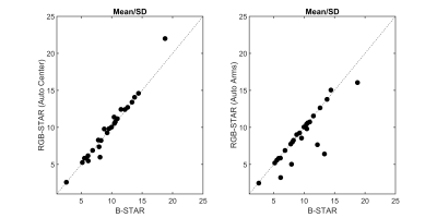
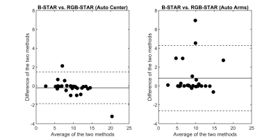
The correlation between the mean/SD values evaluated on 26 test ROIs across two central abdominal MRI slices for 13 subjects using B-STAR and RGB-STAR are shown in Fig. 4. The Pearson’s correlation coefficient,8 r, between B-STAR and the RGB-STAR (Auto Center) method is 0.9837 (p-value: $$$2.2035e^{-19}$$$), and between B-STAR and the RGB-STAR (Auto Arms) method is 0.8694 (p-value: $$$8.2366e^{-9}$$$). Figure 5 shows Bland-Altman plots9 indicating the magnitude of disagreement (both error and bias), outliers, and trends between B-STAR and each of the automated interference covariance matrix generation methods. Thus, both RGB-STAR approaches work well and are comparable to B-STAR as indicated by strong positive correlation in mean/SD values (Fig. 4) and further corroborated by strong agreement in Bland-Altman analysis (Fig. 5).
Conclusion
We propose a seeded region growing image segmentation scheme combined with a beamforming algorithm to automatically generate the interference covariance matrix to suppress streaking artifacts in radial MRI images. Automatic seed point placement for the region growing image segmentation scheme ensures that the entire streaking artifact suppression method is fully automated, thereby reducing tediousness and operator dependence. RGB-STAR yields images with artifact reduction superior to existing methods and is fully automated.Acknowledgements
We would like to acknowledge grant support from NIH (CA245920), the Arizona Biomedical Research Commission (ADHS14-082996), and the Technology and Research Initiative Fund (TRIF).References
1. Xue Y, Yu J, Kang HS, Englander S, Rosen MA, Song HK. “Automatic coil selection for streak artifact reduction in radial MRI.” Magn Reson Med. 2012; 67:470-476.
2. Holme HC, Frahm J. “Sinogram-based coil selection for streak artifact reduction in undersampled radial real-time magnetic resonance imaging.” Quant Imaging Med Surg. 2016; 6:552-556.
3. Walsh DO, Gmitro AF, Marcellin MW. “Adaptive reconstruction of phased array MR imaging.” Magn Reson Med., 2000; 43:682-690.
4. Grimm R, Forman C, Hutter J, Kiefer B, Hornegger J, Block T. “Fast automatic coil selection for radial stack-of-stars GRE imaging.” Proc. ISMRM, 2013, p. 3786.
5. Kholmovski EG, Parker DL, Di Bella EV. “Streak artifact suppression in multi-coil MRI with radial sampling.” Proc. ISMRM, 2007. p. 1902.
6. Mandava S, Keerthivasan MB, Martin DR, Altbach MI, Bilgin A. “Radial streak artifact reduction using phased array beamforming” Magn Reson Med. 2019; 81:3915-3923.
7. Adams R, Bischof L. “Seeded Region Growing”, IEEE T-PAMI, 1994; 16(6):641-647.
8. Kutner MH, Nachtsheim C, Neter J, Li W. “Applied Linear Regression Models.” 4th ed., McGraw-Hill/Irwin, 2004.
9. Altman D, Bland J. “Measurement in medicine: the analysis of method comparison studies,” J R Stat Soc Ser D Stat. 1983; 32:307-317.
Figures
