3513
Aberrant white matter networks in methamphetamine-dependent patients and its application in support vector machine-based classification1Department of Radiology, Ningbo Medical Treatment Center Lihuili Hospital, Ningbo University, Ningbo, China, 2GE Healthcare,Beijing,China, Beijing, China, 3Laboratory of Behavioral Neuroscience, Ningbo Addiction Research and Treatment Center, Ningbo, China, Ningbo, China, 4Ningbo Addiction Research and Treatment Center, Ningbo, China, Ningbo, China
Synopsis
This is a pilot study of the weighted white matter (WM) network in MA-dependent patients. By combining DTI-based probabilistic tractography and graph theory, the WM networks of MA-dependent patients presented small-worldness, and these networks tend to be random networks. The network metrics, that presented inter-group differences were used to construct a support vector machine, that achieved an excellent performance in discriminating MA-dependent patients from normal controls. Overall, the current study demonstrated that MA dependence is associated with abnormal network metrics, and these metrics can be promising features to train a classifier which need further verification with a larger sample size.
Introduction
Methamphetamine (MA) is a highly addictive amphetamine-type psychostimulant. Its excessive consumption causes devastating medical effects and high rates of crime around the world. Brain network is a widely used tool for identifying abnormal topological properties in the human brain1. Abnormal topological metrics provided a new perspective of potential biomarkers for the diagnosis of neurological diseases and had been found in various brain diseases, such as schizophrenia2 and heroin addiction3. However, little is known about the significance of these metrics in the study of MA dependence. Inspired by this, we combined diffusion tensor imaging (DTI)-based probabilistic tractography and graph theory to describe the weighted WM networks. Then we constructed a support vector machine (SVM)-based classifier trained with network metrics to identify MA-dependent patients.Materials and Methods
Forty-six MA-dependent subjects (age: 34.87 ± 7.27, M/F: 46/0) and 46 normal controls (age: 33.13 ± 10.86, M/F: 46/0) were enrolled in the study. The MR examinations were performed on a 3T MRI scanner (DiscoveryTM MR750, GE Healthcare, Milwaukee, WI) equipped with an eight-channel head coil. Conventional axial T2-weighted images were first obtained to rule out cerebral infarction or other lesions. Structural images were acquired using a sagittal 3D T1-weighted sequence (TR/TE = 7.4/3.2 ms; TI = 450 ms; flip angle = 12°; FOV = 256 × 256 mm; matrix = 256 × 256; slice thickness = 1 mm). A single-shot echo-planar imaging sequence in axial plane (TR/TE = 8175/80.8ms; FOV = 256 × 256 mm; matrix = 128 × 128; slice thickness = 2 mm; b-value = 0, 1000 s/mm2) was then used to acquire DTI images without diffusion gradient and with 30 non-collinear diffusion gradient directions.Desterieux and aseg atlas were used to define nodes. The brain was divided into 162 regions, with each region representing a node in the network. FSL software (http://fsl.fmrib.ox.ac.uk/fsl/fslwiki/FSL) was used to analyze the DTI image. The processing steps included: head movement and eddy current correction, removal of brain tissue, estimation of the probability distribution of dispersion direction with bedpostx, and probabilistic tractography between two brain areas by protrackx2 to get the edge of the network.
Data were analyzed using R version 3.4.04. We adopted a network-based statistic approach to assess between-group differences in inter-regional WM connections (p < 0.05, permutation test 1000 times). Area under the curve (AUC) for each network metrics was calculated over the density range and were then compared to test group differences using a general linear model (p < 0.05, permutation test, 10000 times for global metrics and 5000 times for local metrics). Network metrics with inter-group differences were selected to construct a classifier using a linear SVM. A 5-fold cross-validation method, that was repeated for 100 times, was used to evaluate the performance of the model.
Results
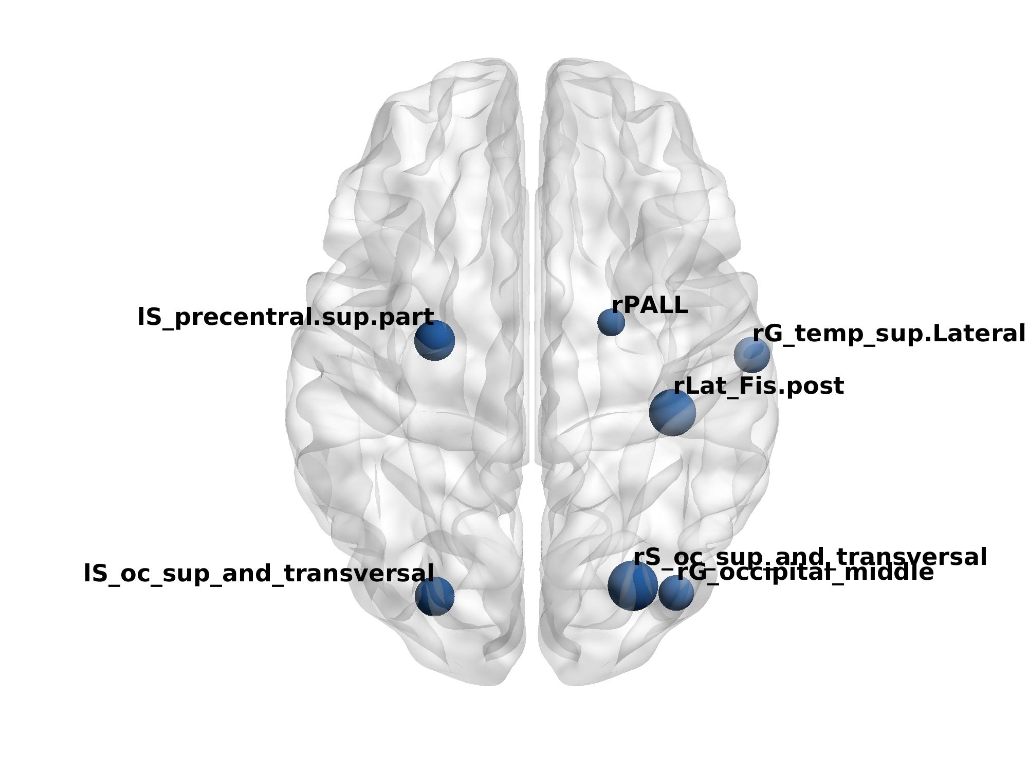
Figure A shows the differences in inter-regional WM connections between MA-dependent patients and the controls. Figure B, C shows the differences in the weighted local efficiency between the MA-dependent patients and the controls. Figure D shows the correlation between weighted local efficiency and the Brief Psychiatric Rating Scale (BPRS) scores. Moreover, the prediction accuracy, sensitivity, specificity, and kappa of 98.86%±2.25%, 99.89%±1.29%, 97.81%±4.38%, and 0.9771±4.50% in differentiating MA-dependents subjects from normal controls were obtained by the classifier, respectivelyDiscussion
The present study showed that MA-dependent patients had a subnetwork characterized by enhanced connection strength, mainly located in the reward system and visual system. It suggested that long-term processing of explicit visual attention stimuli may lead to adaptive changes between two systems, increasing the transmission of information between the two systems, making addicts very sensitive to drug-related cues.Our results confirmed small-worldness in the WM networks of both MA-dependent patients and controls. Compared with the controls, however, the MA-dependent group showed decreased small-world indices and increased clustering coefficient. These results indicated a disturbance between local specialization and global integration in MA-dependent patients. Since the amygdala plays an important role in the control of fear, the increase of the betweenness centrality of the amygdala may be closely related to the increase of negative emotions and stress in interpersonal communication of MA-dependent patients. In addition, the study revealed that brain regions with increased local efficiency are mainly located in the attention system, motivation, and memory circuits, which may be related to the dependents’ long-term attention and learning to the relevant drug stimulation. Interestingly, the local efficiency of most of these brain regions also contributes significantly to the classifier, which also provides machine learning evidence to support that the local efficiency abnormalities of these brain regions are related to MA dependence. Furthermore, local efficiency of the prefrontal cortex, such as the middle frontal gyrus and orbital sulcus, was found to be correlated with BPRS scores. Previous studies also reported functional abnormalities of these brain regions in patients with neuropsychiatric diseases5-6. These results implied that the local efficiency of these brain regions may be one of the neural mechanisms of mental disorders in MA-dependent patients.
Conclusion
The present study provided evidence that MA dependence is associated with abnormal topological metrics, such as disturbed small-worldness and altered local efficiency, which could be used as hopeful features in constructing a SVM-based classifier to diagnose MA-dependent patients with excellent performance.Acknowledgements
The research was partially supported by the funding of National Key R&D Program of China (grant: 2017YFC1310403), the funding of National Basic Research Program of China (grant: 2015CB553504), the funding of National Natural Science Foundation of China (grant: 81471350), the funding of Zhejiang Provincial Medical and Health Science and Technology Program (grant: 2018243996), and the funding of Natural Science Foundation of Ningbo Municipality (grant: 2014A610259, 2017A610224, 2019A610296), and the funding of Science and Technology Program of Ningbo (grant: 202002N3166), and the funding of National Natural Science Foundation of Zhejiang (grant: LGF21H090007).References
[1] Bassett DS, Bullmore ET. Human brain networks in health and disease. Curr Opin Neurol 2009; 22:340–347.
[2] Yu Q, Sui J, Rachakonda S, He H, Pearlson G, Calhoun VD. Altered small-world brain networks in temporal lobe in patients with schizophrenia performing an auditory oddball task. Front Syst Neurosci 2011; 5:7.
[3] Zhang R, Jiang G, Tian J, Qiu Y, Wen X, Zalesky A, Li M, Ma X, Wang J, Li S, Wang T, Li C, Huang R. Abnormal white matter structural networks characterize heroin-dependent individuals: a network analysis. Addict Biol. 2016 May;21(3):667-78. doi: 10.1111/adb.12234. Epub 2015 Mar 4. PMID: 25740690..
[4] R Development Core Team (2011) R: a language and environment for statistical computing. Vienna, Austria: The R Foundation for Statistical Computing
[5] Zhang B, Li S, Zhuo C, Li M, Safron A, Genz A, Qin W, Yu C, Walter M. Altered task-specific deactivation in the default mode network depends on valence in patients with major depressive disorder. J Affect Disord. 2017 Jan 1; 207: 377-383. doi: 10.1016/j.jad.2016.08.042. Epub 2016 Oct 7. PMID: 27750155.
[6] Nishimura Y, Takizawa R, Muroi M, Marumo K, Kinou M, Kasai K. Prefrontal cortex activity during response inhibition associated with excitement symptoms in schizophrenia. Brain Res. 2011 Jan 25; 1370: 194-203. doi: 10.1016/j.brainres.2010.11.003. Epub 2010 Nov 5. PMID: 21059348.
Figures