3477
Banding artifacts reduction for lumbosacral plexus imaging with balanced FFE (b-FFE)1Philips Healthcare, Beijing, China
Synopsis
The balanced FFE (b-FFE) sequence has the advantages of high resolution, SNR, CNR and superior differentiation between fluid and tissue. Therefore, it can be used for lumbosacral plexus imaging [1]. However, black band-like artifacts are frequently observed due to intra-voxel signal dephasing at the presence of magnetic field inhomogeneity. Here we proposed using a RF phase cycling technique (b-FFE-XD) to suppress the banding artifacts. The RF phase was set as [0,180+1*X, 2*X, 180+3*X,4*X,…] in consequence to limit the intravoxel dephasing, where X=n*360/NSA, and n is the nth average. Results demonstrated successful removal of banding artifacts with the proposed technique.
Introduction
The lumbosacral plexus is a complex anatomic area, disfunction of which can lead to severe disability and morbidity [2]. Despite the use of balanced fast field echo (b-FFE) technique, this sequence is prone to magnetic field inhomogeneity and is frequently disturbed by banding artifacts due to intra-voxel signal dephasing with increased severity at 3.0T. This study proposed a RF phase-cycling technique as an addition to the traditional b-FFE sequence (b-FFE-XD) to suppress the potential banding artifacts in lumbosacral plexus imaging.Method
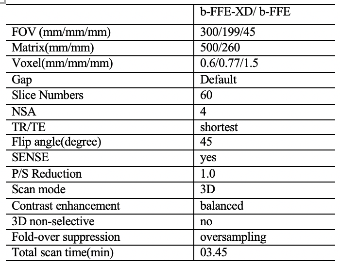
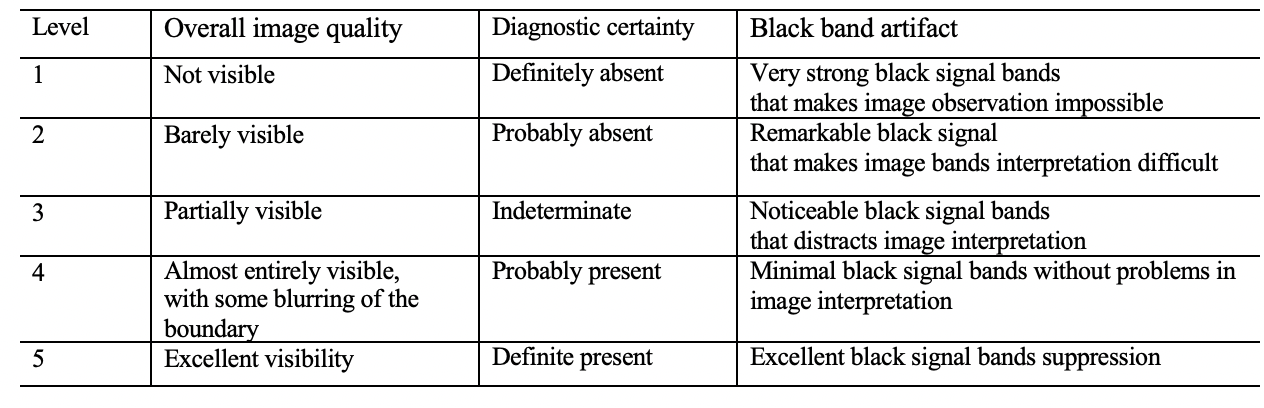
Ten healthy volunteers (4 women, and 6 men, mean age, 53.30±8.73 years; age range, 31-64 years) underwent B-FFE-XD and B-FFE scans (Coronal orientation) at 3.0T (Ingenia Elition, Philips healthcare, Best, the Netherlands) on the lumbosacral plexus with a body coil for RF transmitting and an anterior coil for signal receiving. The detailed acquisition parameters for both sequences were kept the same, listed in Table1 as below, except that a RF phase cycling scheme ([0,180+1*X, 2*X, 180+3*X,4*X,…], where X=n*360/NSA, and n is the nth average) was applied to the excitation pulse for b-FFE-XD. Images were evaluated independently by two radiologists (>3 years radiology experience) in terms of overall image quality, banding artifact suppression, and diagnostic certainty using a 5-point scale criteria (5 for the best result, the scoring system was listed in Table2). Consistency of the evaluation results between the two observers were assessed using intra-class correlation coefficients (ICC) in SPSS (IBM). If the consistency was good, the scores were averaged between the two observers and then used for the subsequent analysis. The scores of the two sequences were compared using a Wilcoxon signed-rank test with a significance threshold of P < 0.05.Results
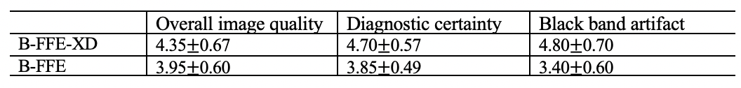
Effective banding artifact suppression was achieved by B-FFE-XD, representative images shown in Figure 1. Good agreement was achieved between the scoring of the two observers, and the ICC of banding reduction was 0.765 (P<0.001). The scores were significantly higher in b-FFE-XD than in b-FFE in all items (overall image quality, diagnostic confidence, black band artifact) under scoring (4.35 0.67 v.s. 3.95 0.60, 4.70 0.57, v.s. 3.85 0.49, 4.80 0.70 v.s. 3.40 0.60, respectively; all P<0.05), as shown in Table 3.Discussion and Conclusion
Our results demonstrated the potential clinical application of the proposed RF phase cycling scheme for lumbosacral plexus imaging, which yielded improved image quality and reduced banding artifacts when compared to the traditional b-FFE technique.Acknowledgements
N/AReferences
[1] F. J. Hans, M. H. Reinges, T. Krings. Lumbar nerve root avulsion following trauma: balanced fast field-echo MRI. Diagnostic neuroradiology. 2004; 46:144-147
[2] Ethan A. Neufeld, Peter Yi Shen, etc. MR imaging of the lumbosacral plexus: a Review of techniques and pathologies. J Neuroimaging. 2015; 25:691-703
Figures
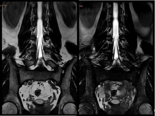
Figure 1.A healthy volunteer, 56-year-old male.
Left: Coronal B-FFE-XD image of the lumbosacral plexus. Right: Coronal B-FFE image of the lumbosacral plexus