3475
Optimization of Compressed SENSE accelerated Brachial Plexus MRI: Quality and Efficiency
Renwang PU1, Qingwei SONG1, Ailian LIU1, Zijing ZHANG1, Nan ZHANG1, Haonan ZHANG1, Bingbing GAO1, Lihua CHEN1, and Liangjie LIN2
1the First Affiliated Hospital of Dalian Medical Universityrsity, Dalian, China, 2Philips Healthcare, BEIJING, China
1the First Affiliated Hospital of Dalian Medical Universityrsity, Dalian, China, 2Philips Healthcare, BEIJING, China
Synopsis
The 3D-NerveVIEW sequence with suppression of lipid and blood signals and a long echo time can obtain high-quality images for visualization of brachial plexus, while the long scan time may limit its clinical application. Compressed SENSE (CS) is a newly developed technique in MRI that enables accelerated acquisition with maintained image quality. By comparing results of 3D-NerveVIEW for brachial plexus imaging with acceleration by the conventional SENSE and the advanced CS with different acceleration factors. We found that the 3D-NerveVIEW for brachial plexus imaging with a CS acceleration factor of 4 can obtained favorable images within significantly reduced scan time.
Synopsis
The 3D-NerveVIEW sequence with suppression of lipid and blood signals and a long echo time can obtain high-quality images for visualization of brachial plexus, while the long scan time may limit its clinical application. Compressed SENSE (CS) is a newly developed technique in MRI that enables accelerated acquisition with maintained image quality. By comparing results of 3D-NerveVIEW for brachial plexus imaging with acceleration by the conventional SENSE and the advanced CS with different acceleration factors. We found that the 3D-NerveVIEW for brachial plexus imaging with a CS acceleration factor of 4 can obtained favorable images within significantly reduced scan time.Introduction
Magnetic resonance imaging (MRI) is the favored modality for evaluating the brachial plexus because of its multiplanar capabilities and excellent soft-tissue contrast. The 3D-NerveVIEW sequence with suppression of lipid and blood signals and a long echo time can obtain high-quality images for brachial plexus, but the long scan time of 3D acquisition can be a problem for clinical applications. Compressed SENSE (CS) is a newly developed technique in MRI that enables acceleration of MR scan without sacrifice of image quality. This study aims to evaluate the application CS on 3D-NerveVIEW imaging for brachial plexus, and to find an optimized acceleration factor.Materials and Methods
This study has been approved by the local IRB. 16 healthy volunteers (7 males, age 22-68, 45.38±13.92) were recruited and underwent the 3D-NerveVIEW scan of brachial plexus on a 3.0 T MR scanner (Ingenia CX, Philips Healthcare, Best, the Netherlands). Scan parameters are as follows: TR: 2200ms, TE: 170ms, FOV: 300X453X96 mm, NSA: 2, voxel size: 1.2X1.2X2.4 mm, matrix: 252X378X?, slice thickness and Gap: 2.4/-1.2 mm, The 3D-NerveVIEW was scanned with different acceleration factors including the conventional SENSE factor of 2 (SENSE2, as reference) and CS factors of 2 to 16 with a interval of 2 (CS2-CS16). 9 sets of data were collected for each volunteer obtained with scan time recorded. The signal intensity (SI) and standard deviation (SD) for brachial plexus and muscle were measured on the AW4.7 workstation (Advantage Workstation, GE Healthcare) for each group on the coronal images. ROIs area were approximately 15 mm2 (Fig.1). SNR and CNR were measured as SNR=SIbrachial plexus/SDmuscle and CNR=(SIbrachial plexus-SImuscle)/SDmuscle. The scan time reduction ratios (STRR = (TimeS2-TIimeCSAF)/TIimeS2) were also calculated. The image quality of each dataset was scored subjectively by two radiologists on a four-point scoring criterion. The SPSS24.0 software was used for statistic analyses. Correlation between acceleration factors and image quality parameters were analyzed by XXX. Shapiro-Wilk test was used to test the normality of quantitative parameters and then the paired-sample t test was used to compare the brachial plexus signal value, background noise, SNR and CNR between the S2 group and the CS groups. The interobserver reliability on qualitative evaluation was assessed via Cohen' s kappa test (excellent agreement if k > 0.9; good agreement if k > 0.6). The Friedman test was used to compare the subjective scores of the images, and the Bonferroni method was used for pairwise comparison.Result
Quantitative parameters conformed to obey normal distribution(P>0.05). Qualitative parameters consistency between the two observers was good (k > 0.6). Brachial plexus signal intensity, SNR, CNR, background noise (SD for muscle tissue) and image quality scores were significantly correlated with CS-AF (all P<0.05). As the AF increases, the brachial plexus signal intensity, SNR, and CNR gradually decreased (r= -0.996, -0.992,-0.992), background noise gradually increased (r= 0.995), and the subjective image score decreased with the increase of AF (r=-0.977)(Fig.1, 2).The brachial plexus SI, SNR, CNR, background noise, image scores of the reference SENSE group were not significantly different from those of the CS2~CS10, CS2~CS4, CS2~CS4, CS2~CS6, and CS2~CS6 groups respectively (all P > 0.05). The scan time of CS2~CS16 was reduced by 9%~88% compared with the SENSE2 group. (Fig.3)Discussion and Conclusion
3D-NerveVIEW scan with a CSAF of 4 is recommended for brachial plexus imaging with significantly reduced scan time (381s vs 821s, 54%) and comparable image quality compared to the reference scan.Acknowledgements
No acknowledgement found.References
[1] Bratke G, Rau R, Weiss K, et al. Accelerated MRI of the Lumbar Spine Using Compressed Sensing: Quality and Efficiency. J Magn Reson Imaging, 2019, 49(7): e164-e175. [2] Lee S H, Lee Y H, Suh J S. Accelerating knee MR imaging: Compressed sensing in isotropic three-dimensional fast spin-echo sequence. Magn Reson Imaging,2018,46:90-97. Bratke G, Rau R, Weiss K, et al. Accelerated MRI of the Lumbar Spine Using Compressed Sensing: Quality and Efficiency. J Magn Reson Imaging,2019, 49(7): e164-e175. [3] Yoneyama M, Takahara T, Kwee TC, Nakamura M, Tabuchi T. Rapid high resolution MR neurography with a diffusion-weighted pre-pulse. Magn Reson Med Sci. 2013; 12(2): 111-119. [4] Sneag DB, Mendapara P, Zhu JC, et al. Prospective respiratory triggering improves high-resolution brachial plexus MRI quality. J Magn Reson Imaging. 2019, 49(6): 1723-1729. [5] Lustig M, Donoho D, Pauly J M. Sparse MRI: The application of compressed sensing for rapid MR imaging. Magn Reson Med, 2007, 58(6): 1182-1195.Figures
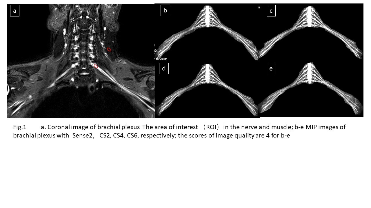
a.Coronal image of brachial plexus.The area of interest(ROI) in the nerve and muscle;b-e.Mip images of brachial plexus of sense2,cs2,cs4,cs6,respectively;the scores of image quality are 4 for b-e.
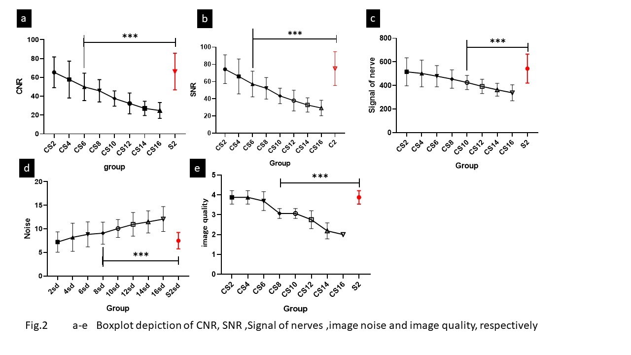
a-e:Boxplot depiction of CNR,signal of nerves,image noise and image quality,respectively.
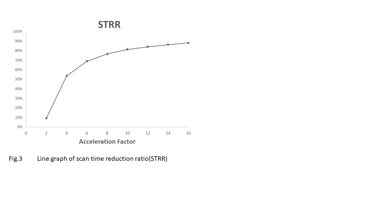
Line graph of scan time reduction ratio(sTRR)