3473
Diffusion features in white and gray matter of healthy cervical spinal cord using PSIR imaging1THE FIRST AFFILIATED HOSPITAL OF DALIAN MEDICAL UNIVERSITY, DALIAN, China, 2Philips Healthcare, BEIJING, China
Synopsis
Due to the difficulty in distinguishing the structure of spinal by conventional MR sequence. In this study, the fusion images of Phase Sensitive Inversion Recovery sequence (PSIR) and zoomed Diffusion Tensor Imaging (DTI) was used to quantify the diffusion indicators, such as fractional anisotropy (FA), apparent diffusion coefficient (ADC), axial dispersion (AD) and relative anisotropy (RA) in the normal spinal cord. We found that there were significant differences of above indicators in different regions of cervical cord including the white columns of the cervical cord, the ipsilateral white matter and in the left and right sides of the cervical cord.
Introduction
The quantitative research on fine structure of gray matter and white matter in the spinal cord is still incomplete [1]. One of the reasons is the fine structure of spinal gray matter and white matter is difficult to be detected by conventional MR sequence. Phase-sensitive T1 inversion recovery images can be acquired by PSIR sequence, which has been used to detect MS lesions both in white and cortical gray matter in the brain and spinal with good tissue contrast [2][3]. Therefore, we aim to explore the diffusion indicators in both ipsilateral and contralateral white and gray matter of the healthy cervical spinal cord.Materials and Methods
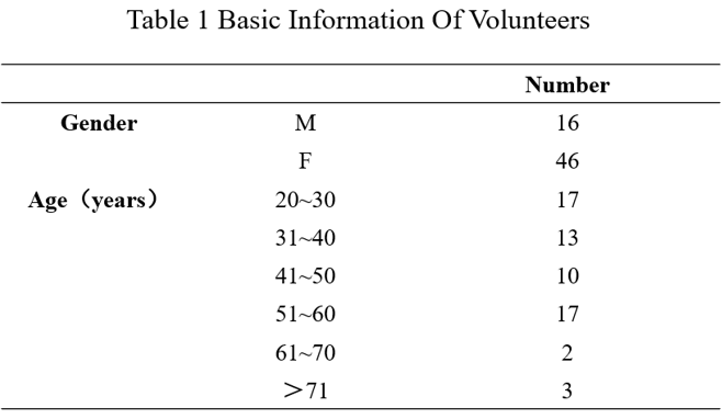
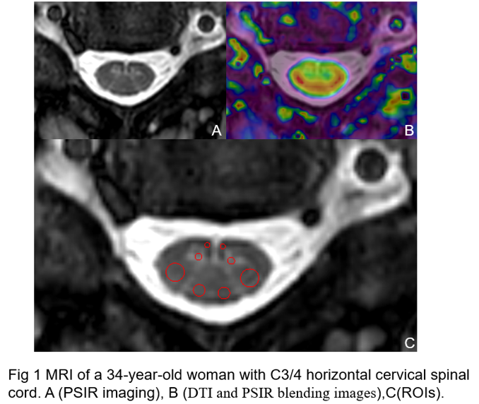
62 healthy volunteers (16 men, 46 women; mean age, 42 years; range, 22–78 years) were recruited in this study (Table 1). Phase Sensitive Inversion Recovery sequence and Diffusion Tensor Imaging were acquired on a 3.0T MR scanner (Ingenia CX, Philips Healthcare, the Netherlands) with a 32-channel head coil.DTI-ZOOM imaging was performed using a single excitation plane echo sequence and its imaging parameters were: TE/TR = 66ms/3.0s, FOV = 220*220 mm2, voxel size = 2.50/0.71/2mm, slice thickness and gap= 2.0 mm/0.0mm, b factor=0 s/mm2 and 800 s/mm2, direction of diffusion sensitive gradient=12 and scan duration=6 min 15s; PSIR imaging was performed using turbo spin echo sequence and its imaging parameters were as follows: TE/TR =9ms/4.0s, FOV = 120mm, voxel size = 0.55/0.63/3.00mm, slice thickness and gap= 3.0 mm/0.0mm, scan duration=2 min 40s. ROIs were drawn on the fusion images of DTI and PSIR by one observer. Eight ROIs were placed on the bilateral anterior horn of gray matter and anterior, middle, and posterior columns of bilateral white matter (Fig.1) [1]. The mean value of DTI parameters (FA, ADC, AD, RA) in those ROIs was recorded.
SPSS was used to analyze the differences of DTI parameters in both ipsilateral and contralateral white and gray matter of spinal. Kolmogorov-sminov test was used for normal test, and wilkerson symbolic rank test was used to test the possible differences.
Results
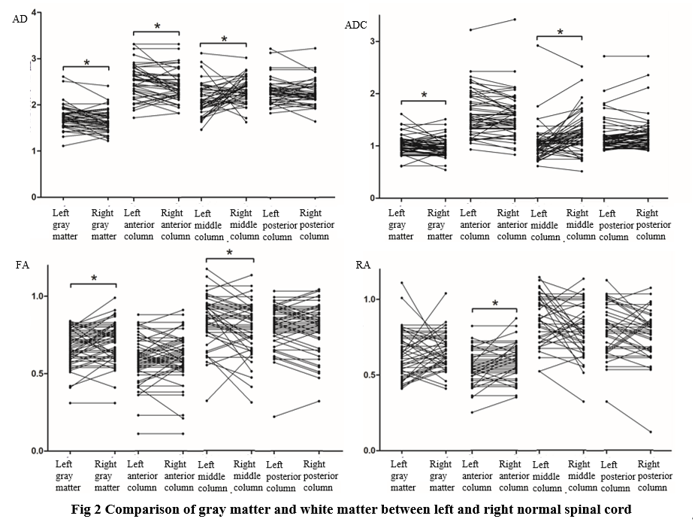
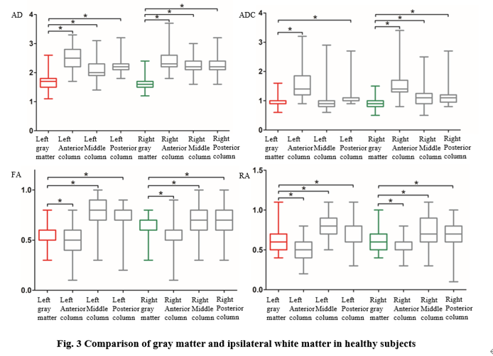
As for the comparation among ipsilateral and contralateral gray and white matter in normal cervical spinal, there are statistical differences in DTI parameters (FA, ADC, AD, RA) (P<0.05) . Compared with the diffusion indicators in contralateral gray and white matter, increased FA was found in right gray matter and left white matter with statistical differences (P<0.05); increased RA was found in right anterior column with statistical differences (P<0.05);decreased ADC and AD were found in right gray matter with statistical differences (P<0.05); decreased AD was also found in right anterior and left middle column with statistical differences (P<0.05); (Fig.2).Compared with the diffusion indicators in bilateral gray matter, increased ADC,RA,AD and FA were found in bilateral white matter with statistical differences (P<0.05); decreased FA and RA were found in bilateral anterior columns with statistical differences (P<0.05); (Fig.3)Discussion
This study reports that there are diffusion differences in bilateral and ipsilateral gray and white matter of healthy spinal cords. The reasons of this results maybe related with right-handedness [3]. Further studies with larger samples need to perform to explore the difference based on the principle of DTI imaging and assess its possible clinical values in patients with spinal disease [4].Conclusion
We found that diffusion differences exist in bilateral and ipsilateral gray and white matter of healthy spinal cords. In spinal cord diffusion quantification studies, this differences should be taken into consideration.Acknowledgements
This abstract was completed under the careful guidance of my mentor Professor Miao Yanwei. I was greatly helped, encouraged and patiently guided by the topic selection, thesis proposal, design and the final completion of the paper.Here to express the most sincere thanks and blessing to my teacher.References
[1] Li DC, Malcolm JG, Rindler RS, et al. The role of diffusion tensor imaging in spinal pathology: A review. Neurol India. 2017. 65(5): 982-992.
[2] Mirafzal S, Goujon A, Deschamps R, et al. 3D PSIR MRI at 3 Tesla improves detection of spinal cord lesions in multiple sclerosis. J Neurol. 2020. 267(2): 406-414.
[3] Liu M, Liu S, Ghassaban K, et al. Assessing global and regional iron content in deep gray matter as a function of age using susceptibility mapping. J Magn Reson Imaging. 2016. 44(1): 59-71.
[4] Zhao M, Shi B, Chen T, et al. Axial MR diffusion tensor imaging and tractography in clinical diagnosed and pathology confirmed cervical spinal cord astrocytoma. J Neurol Sci. 2017. 375: 43-51.
Figures