3471
Visible “Butterfly” of the Cervical Spinal Cord: A Pilot study using high-resolution Phase-Sensitive and multiple Fast Field Echo MR imaging
bingbing gao1, Yanwei Miao1, Qingwei Song1, Ailian Liu1, and Jiazheng Wang2
1the First Affiliated hospital of Dalian Medical university, Dalian, China, 2Philips Healthcare, BeiJing, China
1the First Affiliated hospital of Dalian Medical university, Dalian, China, 2Philips Healthcare, BeiJing, China
Synopsis
Discrimination of gray and white matters on the axial-spinal-marrow MRI remains a challenge, due to the small structure size and motion artifacts from such as swallowing and blood flow. The present study aims to visualize the cervical spinal marrow with high resolution axial phase-sensitive inversion recovery (PSIR) and multiple Fast Field Echo (FFE) images, and further to detect the image quality differentially on gray and white matters. The “butterfly” structure could be seen on both of the two contrast images, but the PSIR images were associated with significantly higher signal-to-noise ratios.
Introduction
Recently, there has been some studies about diseases involving the spinal cord published1,2, among them, the lesions were mostly detected on the sagittal images using MR sequences such as phase-sensitive inversion recovery (PSIR) and multiple Fast Field Echo (FFE), etc. The exact lesion location in an axial image of spinal marrow may imply different diagnosis3. Moreover, detection of lesions locating inside gray or matter of spinal cord can increase the confidence in clinical diagnoses of MS or can predict the conversion of a clinically isolated syndrome into definite MS4. While discrimination of gray and white matters on axial-spinal-marrow MRI remains a challenge, due to the small structure size and motion artifacts from such as swallowing and blood flow. Therefore, here we aim to explore the feasibility of high-resolution axial PSIR and multiple FFE for differential imaging of gray matter (GM) and white matter (WM) in cervical spinal cord.Methods
We recruited sixteen healthy subjects (mean age 28.9±2.36 years; nine males). All subjects underwent MR examinations at a 3.0 T scanner (Ingenia CX, Philips Healthcare, the Netherlands) with a 32-channel neck-head array coil. PSIR and multiple FFE scans were performed at the superior cervical spinal cord (segments C1 to C4, parameters listed in Table 1). Signal intensities of GM, WM and cerebral-spinal-fluid (SI-GM, SI-WM, and SI-CSF) were measured on segments C1 to C3 by two radiologists. The GM-to-WM contrast ratio, GM-to-CSF and WM-to-CSF contrast to noise ratios (CR-G/W, CNR-G/C, and CNR-W/C) and signal-to-noise ratios (SNR-G/C and SNR-W/C) were calculated for the two type images of all subjects. Inter-reader agreement was evaluated using the intraclass correlation coefficient (ICC), and the mean values by the two observers were taken for further analyses. Differences between SI-GM and SI-WM were evaluated with test for both sequences, and differences of CR, SNR and CNR values between the two sequences were assessed using the Mann-Whitney U test.Results
Measurements between the two radiologists were in good agreement (Table 2, 0.713 < ICC < 0.943 for all measurements). The SI-GM is significantly higher the SI-WM in the PSIR images (Table 3, P=0.002), while there was no significant difference between SI-GM and SI-WM in multiple FFE images (Table 3, P=0.125). The CR-G/W values measured from PSIR images were significantly greater than those from multiple FFE Images. The multiple FFE showed significantly greater SNR than PSIR for both GM (P=0.000) and WM (P=0.000). No significant difference was found for CNR-G/C (P = 0.874) and CNR-W/C (P = 0.603) between two sequences (Table 4).Conclusion
In summary, high-resolution PSIR and multiple FFE were used for axial imaging of cervical spinal cord, and the boundary between gray and white matter was observed. The high-resolution PSIR is superior to multiple FFE in differential imaging of GM and WM in the cervical spinal cord with an acceptable SNR reduction.Acknowledgements
The authors are grateful to the Department of Radiology, First Affiliated Hospital of Dalian Medical University, for supporting this study. The authors thank all volunteers who participated in this study.References
1. Fechner A, Savatovsky J, El Methni J, et al. A 3T Phase-sensitive inversion recovery MRI qequence improves detection of cervical spinal cord lesions and shows active lesions in patients with multiple sclerosis. AJNR Am J Neuroradiol. 2019;40(2):370-375. 2. Nelson F, Poonawalla AH, Hou P, et al. Improved identification of intracortical lesions in multiple sclerosis with phase-sensitive inversion recovery in combination with fast double inversion recovery MR imaging. AJNR Am J Neuroradiol. 2007;28(9):1645-9.Figures

Table 1. scan parameters of PSIR and multiple FFE

Table
2. ICC between two observers in GM and WM signal intensity of PSIR and FFE

Table 3.
comparison of GM to WM signal intensity measured
from the high resolution PSIR and FFE

Table
4. comparisons of CNR and SNR values measured from the high resolution
PSIR and FFE images
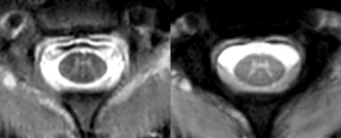
Figure1 Axial cervical spinal
cord PSIR (a) and multiple FFE (b) images