3468
Partial Simultaneous Multi-Slice Acquisition of Combined T2*-Weighted Imaging of the Human Brain and Cervical Spinal Cord1Department of Systems Neuroscience, University Medical Center Hamburg-Eppendorf, Hamburg, Germany
Synopsis
Combined fMRI T2*-weighted imaging of the human brain and cervical spinal cord has been accelerated by simultaneous multi-slice (SMS) imaging of the brain volume. The implementation has been tested and evaluated in phantoms and in vivo and shows a very similar performance to non-accelerated and brain-only measurements. With the much shorter acquisition times achievable it could help to improve brain volume coverage of cortico-spinal fMRI.
Introduction
Combined functional magnetic resonance imaging (fMRI) of the brain and cervical spinal cord1 allows to assess the functional connectivity between the two regions, e.g., in pain processing2 or motor execution. However, volume coverage and temporal resolution were limited because simultaneous multi-slice (SMS)3 imaging could not be used for such measurements due to the two different slice groups involved and the low SMS performance of typical neck coils used for the spinal cord slices. Here, a partial-SMS acceleration for the brain volume has been implemented and tested that may help to overcome these limitations.Method
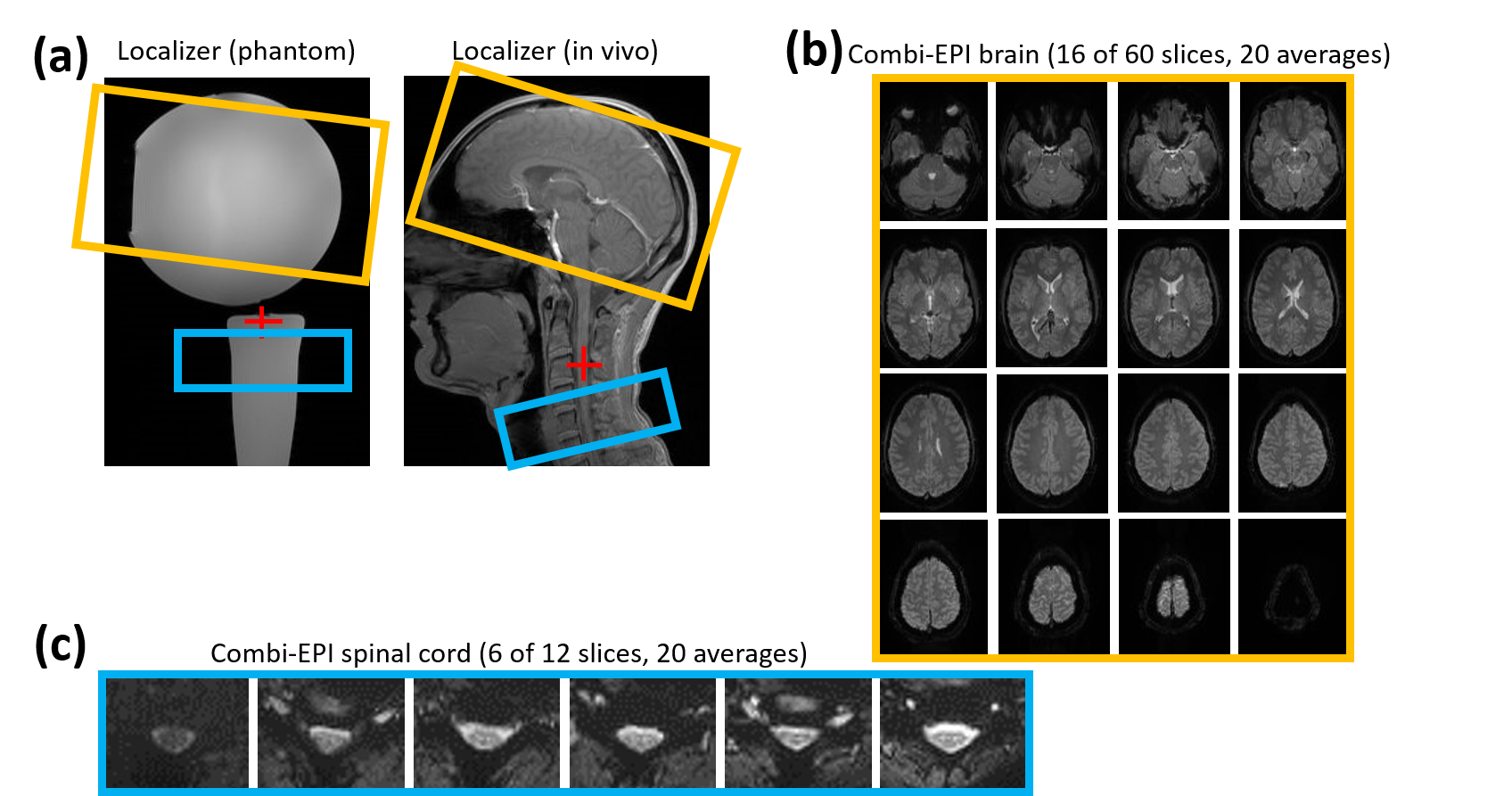
Experiments were performed on a 3T whole-body MR system (PrismaFit, Siemens Healthineers) using a 64-channel head-neck coil with a spherical and a flat water phantom and in vivo. Healthy volunteers were investigated after their informed consent was obtained. EPI acquisitions of the brain volume (60 slices) were performed using a square FOV of 220mm, an in-plane resolution of 2.0×2.0mm2, and a slice thickness of 2mm without gap; for the spinal cord volume (12 slices), the FOV was 136mm, the in-plane resolution 1.2x1.2mm2, and the slice thickness 3.5mm without a gap (cf. Fig. 1). 7/8 partial Fourier and GRAPPA with an acceleration factor of 2 and 36 reference lines was used for both volumes. The brain and spinal cord volumes were acquired with a TE of 24ms and 27ms, respectively, yielding a minimum TR of 4177ms without SMS acceleration, 2524ms with brain SMS for an acceleration factor of 2, and 1975ms with brain SMS for an acceleration factor of 3. The minimum TRs with a flip angle of 75° were used for the water phantom measurements. In vivo, a fixed TR of 4177ms with a flip angle of 86° was used for all acquisitions.A dynamic update of the frequency and first order shim values was performed between the two volumes as described previously1 for all acquisitions. The isocenter was positioned close to the spinal cord volume (see Fig. 1a) which improved the overall shim.
The image reconstruction pipeline was modified to ignore the SMS part of the reconstruction for spinal cord slices while brain slices passed the full pipeline including the SMS functionality.
To evaluate the performance of the partial SMS acceleration in the brain, the images were compared to data obtained for the brain volume only with the same SMS acceleration factors.
Results
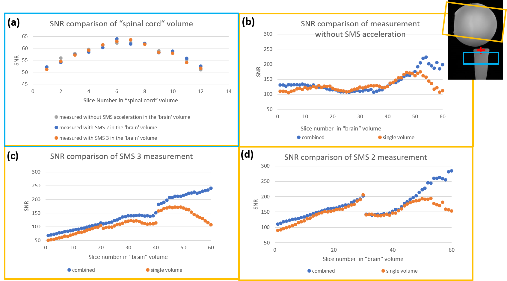
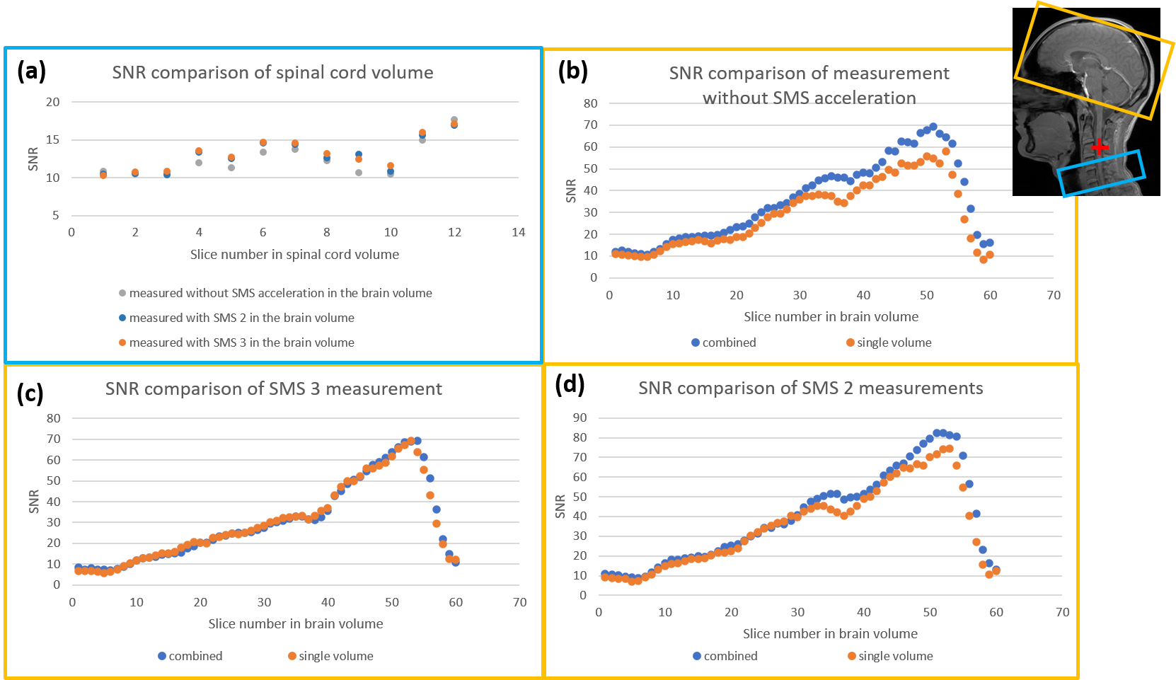
The localizers and brain and spinal cord example EPI images are shown in Figure 1, the latter two were obtained in a single measurement covering both volumes ("combined") with an SMS acceleration factor of 3 for the brain volume.Figures 2 and 3 summarize results of SNR measurements in the phantoms and in vivo, respectively. No impact of the SMS acceleration of the brain is observed in the spinal cord (Fig. 2a and 3a) indicating that the SMS acquisition and reconstruction for the brain slices does not interfere with the non-accelerated spinal cord slices and the chosen reconstruction bypass for the spinal cord slices only opts out SMS functionality. Comparing single-volume and "combined" measurements for brain slices reveals no relevant difference in lower slices, neither without, nor with SMS acceleration which indicates that the partial acceleration of the "combined" measurements provides the same performance as a full-SMS single volume acquisition. This also holds true for upper slices in vivo; only in the phantom experiments, a difference is observed in upper slices which, however, is related to coil elements that were inactive during the single-volume acquisitions.
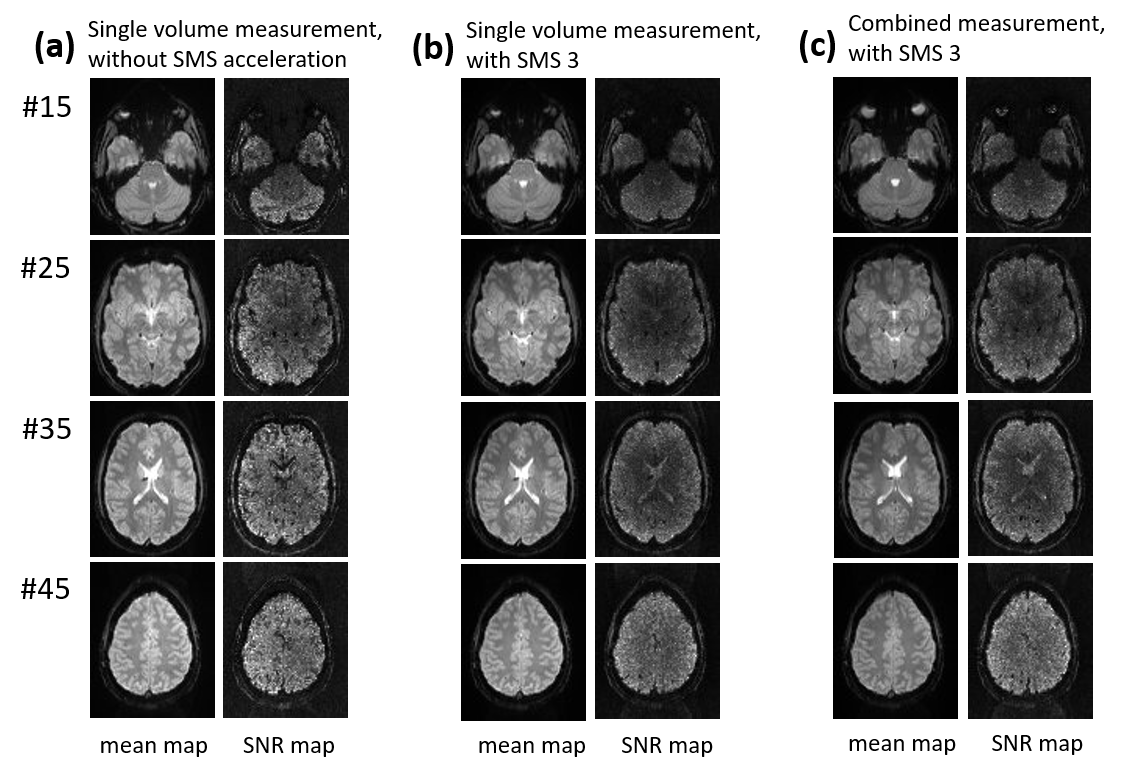
MR images and SNR maps of several slices in the brain volume are shown in Fig. 4, which shows that combined fMRI with an SMS factor of 3 for the brain volume has a very good image quality that is comparable to that of a single-volume acquisition, both without and with SMS. However, the minimum TR achievable is reduced from 4177ms to 1975ms. The SNR maps even seem to indicate a better SNR with SMS which, most likely, is related to noise filtering performed during the SMS reconstruction.
Conclusion
In this study, simultaneous multi-slice acceleration of combined T2*-weighted acquisitions of the human brain and cervical spinal cord was implemented and tested for the brain volume ("partial-SMS"). The results show a similar image quality compared to both single-volume measurements covering the brain only and non-accelerated combined acquisitions and demonstrate the good performance of the implemented approach. With the full image reconstruction being performed on-the-fly and a reduction of the image acquisition time by more than 50%, it may help to improve the temporal resolution and/or brain volume coverage of cortico-spinal fMRI.Acknowledgements
This research was supported by the German Research Foundation DFG (SFB936/A6).References
1. Finsterbusch J, Sprenger C, Buchel C. Combined T2*-weighted measurements of the human brain and cervical spinal cord with a dynamic shim update. NeuroImage.2013;79:153-161.
2. Tinnermann A, Geuter S, Sprenger C, Finsterbusch J, Büchel C. Interactions between brain and spinal cord mediate value effects in nocebo hyperalgesia. Science.2017;358:105-108.
3. Barth M, Breuer F, Koopmans P, Norris D, Poser B. Simultaneous multislice (SMS) imaging techniques. Magn Reson Med.2015;75(1):63-81
Figures