3453
Lumbar disc degeneration changes in sedentary and prolonged-standing population assessed by T1ρ and T2 mapping Magnetic Resonance Imaging
Zeng qi1, Zhang ziwei1, Nie lisha2, Zhu xia1, Huang zaoshu1, and Song lingling1
1The affiliated hospital of Guizhou mdeical university, Guizhou guiyang, China, 2GE Healthcare,MR Resertch China, Beijing, China
1The affiliated hospital of Guizhou mdeical university, Guizhou guiyang, China, 2GE Healthcare,MR Resertch China, Beijing, China
Synopsis
Lumbar disc degeneration is an important cause of lower back pain, sedentary, prolonged-standing behavior accelerates its course. In this study we evaluate intervertebral disc degeneration quantified by T1ρ and T2 mapping magnetic resonance imaging in sedentary and long standing populations compared with a healthy control group matched for age. We conclude that prolonged-standing is more likely to affect the L1 / 2 level discs, while sedentary is more likely to affect the lower segment discs (L4 / 5 and L5/S1).
Introduction
Low back pain is one of the most common clinical diseases, which affects approximately 637 million people worldwide [1]. Lumbar intervertebral disc degeneration (IVDD) is an important cause of low back pain. IVDD typically begins at 20 years of age and excessive mechanical loading (Sedentary, prolonged-standing behavior and high impact loads) accelerates its course [2]. Magnetic resonance imaging (MRI) is a non-invasive medical imaging modality that provides excellent anatomic detail of spinal tissues, and Pfirrmann et al devised a five grade grading system based on MR signal intensity, disc structure, nucleus to annulus distinction and disc height [3]. However, this scoring system is limited in the evaluation of IVDD due to the unsatisfactory objectivity. Quantitative MRI, such as T1ρ、T2 mapping technique, can reflect the molecular environment in early degeneration discs and provide objective continuous measures[4]. However, there are limited studies comparing quantitative IVD measures of sedentary and prolonged- standing behavior individuals. This study aimed to evaluate the performance of T1ρand T2 mapping in disc degeneration assessment in sedentary and prolonged-standing populations.Methods
Subjects: 30 volunteers (14 sedentary, 8 prolonged-standing, 8 healthy volunteer; age range from 23 to 44 years; BMI range: 21.9-24.8) were included in this study with Institution Review Broad approval. Inclusion criteria consisted of: (1)no contraindications for MR examination; (2) no obvious scoliosis or spondylolisthesis; (3)no history of spine trauma and deformity; (4) no metabolic or autoimmune diseases; (5)the sedentary group and the prolonged-standing group spent an average of more than 6 hours per day ; (6)the healthy volunteer have no history of vigorous exercise for long periods; (7) BMI<25 kg /m2. MR scanning was performed using a GE Discovery 750W 3.0T scanner with a spine coil. The key protocols were performed in the table1.For measurement, the disc was divided into five equal areas, the first covering the anterior annulus fibrosus (AAF), the middle covering the nucleus pulposus (NP), and the last covering the posterior annulus fibrosus (PAF), mean values of regions of interest (ROI, 5mm diameter) were measured. In addition, two clinical radiologist using the system described by Pfirrmann examined and graded midsagittal sections of MRI T2 weighted images of 5 lumbar discs. Statistical methods:Statistical analyses were conducted using SPSS 19.0 software. Inter-and intra‑observer reliabilities of Pfirrmann grading were examined using the kappa concordance test. We compared median T1ρ、T2 values of NPs and AFs of healthy volunteers, sedentary, and prolonged-standing groups according to the anatomic level of each disc, using the Kruskal Wallis test. Scatter plots were mapped and the Spearman rank correlation coefficient were applied to determine the relationship between NP、AF and Pfirrmann grade, and the decreasing trend of the relationship between NP、AF and age were evaluated as well. P < 0.05 was considered statistically significant.Results
The T1ρ values of 145 discs, T2 values of 150 discs were successfully obtained eventually. MRI indicated that the T1ρ values of NP at L1/2 were significantly reduced in the prolonged-standing group, whereas there was no significant difference between the sedentary group and the healthy volunteers. The T1ρ values of NP at L4/5 and L5/S1 discs were significantly and mildly reduced in the sedentary group and the prolonged-standing group respectively. In addition, the T2 values of NP and PAF at L4/5 and L5/S1 discs were significantly and mildly reduced in the sedentary group and the prolonged-standing group respectively. For NP and AF, the T1ρ and T2 values were significantly different between the Pfirrmann grades (P<0.05; with the exception of grade I and grade II、grades IV and V). The T1ρ and T2 values decreased with the increase of age, there was no statistically significant difference between the T1ρ and T2 values.Discussion and Conclusion
This study quantitatively assessed the disc degeneration in sedentary and prolonged-standing populations with T2 mapping and T1rho. The previous literatures suggest that behaviors such as spine overload (sedentary, prolonged standing) may be detrimental to the disc and leading cause of catabolism [2]. Our results suggest that sedentary behavior and prolonged-standing behavior can affect different levels of the lumbar disc. Prolonged-standing is more likely to affect the L1 / 2 level discs, while sedentary is more likely to affect the lower segment discs (L4 / 5 and L5/S1). The different target spot in each vertebral level with certain movements may explain why segment of discs varies between different overload [5]. However, spine overload factors cannot be considered as the only cause of degeneration. IVDD is also influenced by the increase of age, and the lower segment discs are inherently more susceptible to degeneration than upper segments [6]. In addition, we found that significant Pfirrmann grades-related reduction of T1ρ and T2 values, however, the detection efficacy was roughly comparable between T1ρ and T2 mapping. The results can be considered as preliminary given the small sample size. The T1ρ and T2 mapping are promising techniques for the diagnosis of IVDD, and may be a useful guide to evaluate patients in future clinical trials of IVD regeneration strategies [7].Acknowledgements
No acknowledgement found.References
[1] Global, regional, and national incidence, prevalence, and years lived with disability for 354 diseases and injuries for 195 countries and territories, 1990-2017: a systematic analysis for the Global Burden of Disease Study 2017. Lancet. 2018. 392(10159): 1789-1858. [2] Belavý DL, Albracht K, Bruggemann GP, Vergroesen PP, van Dieën JH. Can Exercise Positively Influence the Intervertebral Disc. Sports Med. 2016. 46(4): 473-85. [3] Pfirrmann CW, Metzdorf A, Zanetti M, Hodler J, Boos N. Magnetic resonance classification of lumbar intervertebral disc degeneration. Spine (Phila Pa 1976). 2001. 26(17): 1873-8. [4] Iriondo C, Pedoia V, Majumdar S. Lumbar intervertebral disc characterization through quantitative MRI analysis: An automatic voxel-based relaxometry approach. Magn Reson Med. 2020. 84(3): 1376-1390. [5] Vadalà G, Russo F, Battisti S, et al. Early intervertebral disc degeneration changes in asymptomatic weightlifters assessed by t1ρ-magnetic resonance imaging. Spine (Phila Pa 1976). 2014. 39(22): 1881-6. [6] Wang YX, Griffith JF, Leung JC, Yuan J. Age related reduction of T1rho and T2 magnetic resonance relaxation times of lumbar intervertebral disc. Quant Imaging Med Surg. 2014. 4(4): 259-64. [7] Rosenzweig DH, Fairag R, Mathieu AP, et al. Thermoreversible hyaluronan-hydrogel and autologous nucleus pulposus cell delivery regenerates human intervertebral discs in an ex vivo, physiological organ culture model. Eur Cell Mater. 2018. 36: 200-217.Figures
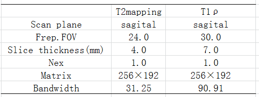
Table 1.The key protocols,GE Discovery 750W 3.0T
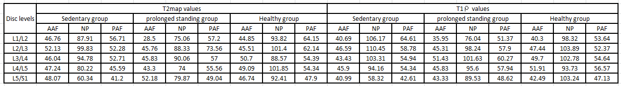
Table 2. T1、T2 values of NP、AAF、PAF according to the anatomic level of each disc NP:nucleus
pulposus; AAF:anterior annulus
fibrosis; PAF:posterior annulus fibrosis
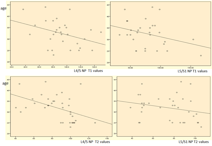
Figure 1. L4/L5、L5/S1 NP values were negatively correlated with age
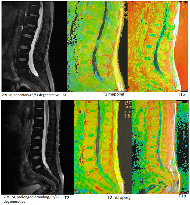
Figure2. Two 29-year-old men, prolonged-standing affected L1/2 disc, sedentary affected L5/S1 disc, the nucleus pulposus was poorly demarcated from the annulus fibrosus